|
Molly A. Schnirring-Judge, DPM, FACFAS - Director of Podiatric Clerkship Program
- Department of Surgery
- St. Vincent Charity Hospital
- Cleveland, Ohio
Robaxin dosages: 500 mg
Robaxin packs: 60 pills, 90 pills, 120 pills, 180 pills, 270 pills, 360 pills

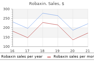
Best buy for robaxinIn recent years much has been understood concerning the molecular mechanisms of ectodermal embryogenesis and this has allowed the establishment of a more rational basis for the classification of ectodermal dys plasia spasms while eating order cheap robaxin on line. Interestingly, not only fullblown ectodermal syndromes but also mono or oligosymptomatic ectodermal malformations might outcome from a mutation in an ectodermal key gene. Embryo genesis happens in distinct tissue organizational fields and specific interactions among the many germ layers which may lead to a wide range of ectodermal dysplasias. The pores and skin is the product of ectodermal and mesodermal stem cell differentiation [11]. Orchestrated pores and skin improvement is simply possible by intensive data trade particularly between the meso derm and ectoderm. Mesodermal signalling pathways corresponding to wingless (Wnt) are essential for the induction of appendages of the pores and skin. Wnt signalling, for example, has influences on mes enchymal cells and on epithelial cells [13]. Whereas the mesoderm induces the placodes by Wnt, the ectoderm evolves into epi dermis with the assistance of ectodysplasin and sonic hedgehog [14]. There is a crosstalk of a number of signalling cascades, such because the ectodysplasin and p53 signalling pathways. Genetic defects in signalling pathways, which disturb the interplay between the ectoderm and mesoderm, result in ectodermal dysplasia. Dickkopf expression results in the place and orientation of the placode and the sonic hedgehog cascade is liable for the formation of hair germ. Ripply, which is a retinoic acidinducible repressor, is required for setting the borders of the preplacodal ectoderm [15]. Disease embryo development network analysis reveals a relationship between disease genes and embryo devel opment�associated genes [17]. Ectodermal dysplasias are noticed globally and this prevalence might apply to quite a few places in the world [10]. Age the diagnosis of ectodermal dysplasias are made inside the first years of life, typically after issues corresponding to hyperthermia or pneumonia have occurred. The role of Wnt in physiology and illness is properly documented in quite a few papers [20]. In the skin, Wnt sign ling is important for the establishment of the dermatome archi tecture, dermis, skin areas with particular person appendages, axial determination and intraappendage regions [21]. Also the uncommon syndrome of odontoonychodermal dysplasia is attributable to mutations in the identical gene. Because p63 has so many ligands and is linked with many different signalling path methods, the medical options of p63 illnesses are rather heterogeneous and broad [26]. Nonsyndromic cleft lip has been observed with mutations in p63 [28], which underlines the fact that monosymptomatic ectodermal malformations could result from a mutation in an ectodermal key gene. Signalling pathways in human skeletal dysplasias usually additionally have an effect on the pores and skin. Numerous mutations in genes that encode for adhesion molecules can result in ectodermal dysplasia, includ ing mutations in connexin genes causing keratitis�ichthyosis� deafness syndrome, oculodentodigital dysplasias, Clouston syndrome, Vohwinkel syndrome, Bart�Pumphrey syndrome and others. In addition mutations in des mosomeforming genes can even result in combined pores and skin and automobile diac issues. There are numerous illnesses with mutations in genes necessary for cell to cell adhesion that are placed within the ectodermal dysplasias however with out clear systematics [32]. The following part evaluations some of the commoner ectoder mal dysplasias, classified in accordance with the signalling pathway concerned in their pathogenesis. In addition, this transcription issue performs crucial roles in the improvement and homeostasis of the epidermis and the correct perform of lymphatic vessels [34]. In many instances these predominantly phenotypedriven, mouse�human comparability research have yielded vital new insights into molecular pathways. The crinkled mouse (cr) is a spontaneous mouse mutant with a phenotype similar to downless and tabby [18]. Because p63 is linked with many different signalling pathways, the clinical features of p63related diseases is somewhat heterogeneous and broad [26]. Numerous genes are differentially expressed in the absence of a fully useful p63. Epigenetic modifications have been proven to affect p63 regulation of terminal differentiation genes. The E3 ligase itch mediates the degradation of p63, providing another degree of regulation and thus suggesting novel therapeutic strate gies for ectodermal dysplasia [47]. The p53 gene household contains key regulators of the cell cycle, that are mutated in additional than 50% of human cancers. They regu late the development of facial prominence and limb buds, and are important for cranial closure and development of the lens [26]. Limb defects are greatest explained by a failure of the apical ectoder mal ridge to develop [48]. Some genotype�pheno sort correlations have been identified (for a evaluate see [49]. A subsequent report has demonstrated a mutation that confers significant transactiva tion exercise on Np63, an otherwise inert isoform of p63 [49]. This autosomal dominant syndrome was first reported in a Dutch family with a constellation of features that had not previously been reported, including hand and foot anomalies and mammary gland aplasia/hypoplasia. The pores and skin and hair were regular in all affected individuals, but some had lacrimal duct atre sia, nail dysplasia, hypohidrosis, hypodontia or cleft palate [49] In addition, inner feminine genital dysplasias have been noticed [53]. Defects in transcription components aside from p63 In addition to the p63 pathway, a quantity of different ectodermal dyspla sias have now been attributed to defects in transcription components that management the expression of a quantity of goal genes important in ectodermal morphogenesis [54]. In many circumstances, positional clon ing studies have yielded the primary mutation in a specific gene however the detailed molecular signalling pathways have yet to be delineated. There is evidence that the 2 ciliary proteins, Evc and Evc2, the merchandise of human dis ease genes associated with the EvC syndrome, act downstream of smoothened to transduce sonic hedgehog signalling. A third baby had a milder phenotype with repeated infections because of Staphylococcus aureus and Streptococcus pneumoniae, and three further siblings from a special kindred had recurrent severe infections with S. Since these unique reviews, the disease phenotypic spectrum has been delineated and consists of extreme lifethreatening or recurrent bacte rial infections within the lower respiratory tract, pores and skin, gentle tissues, bones and gastrointestinal tract, as properly as meningitis and septicaemia in early childhood. Most patients have extreme hypogamma globulinaemia, with low serum IgG levels and various levels of different immunoglobulin isotypes (IgA, IgM and IgE). Some patients have massively elevated IgM ranges, and an impaired antibody response to polysaccharides is essentially the most consistent characteristic of this situation. It has been proven that the pathogenic mutation preferentially impairs the interaction with K63 and M1linked diUb, which correlates with its ubiqui tinbinding defect in vivo [5]. Genetics As ectodermal dysplasias are such a heterogeneous group of illnesses, all inheritance patterns have been described includ ing autosomal recessive, autosomal dominant, Xlinked and mitochondrial. Environmental components Epigenetic factors seem to be important for the phenotypical expression of the disease. Often sufferers have comorbidity with inflammatory bowel illness and rheu matoid arthritis.
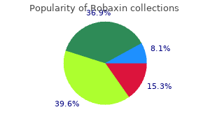
Robaxin 500 mg low costThe significance of noninfectious inflammatory problems is more and more recognized muscle relaxant generic discount 500 mg robaxin with amex. These include inflammatory bowel illness, which clinically and histologically can be indistinguishable from Crohn illness, restrictive lung defects, acute genitourinary obstruction and cutaneous granulomata particularly at vaccination websites. The illness has numerous clinical manifestations, but the hallmark is acute, and potentially fatal, bacterial or fungal an infection. Patients endure from organ abscesses with staphylococci or fungi within the spleen, liver and lung, as well as lymph node abscesses. Any space the place the skin has been broken, by abrasion for example, tends to turn into impetiginized or ecthymatous. Nodular lesions may follow, and these incessantly break down to type necrotic ulcers. Subcutaneous nodules could develop at immunization sites, and these additionally tend in time to ulcerate. Poor healing of surgical wounds, and of the discharging nodular pores and skin lesions, Defects of neutrophil differentiation There are no less than 9 subtypes of congenital neutropenia which were described. Patients with neutropenia present normally in infancy when omphalitis, pores and skin and liver abscesses are most common. Moreover, there may be oral ulceration, gingivitis and early loss of permanent enamel. Patients have aggressive periodontitis, leading to dental loss and palmoplantar hyperkeratosis. Patients current with poor wound healing despite marked leucocytosis in their blood. The scientific image is almost completely explained by the method in which by which leukocytes are drawn to areas of infection and irritation. This leads to blockage of small vessels and quickly expanding necrotic lesions without pus. Gingivitis, ulcerative stomatitis and periodontitis are frequent and severe, leading to loss of teeth. Inflammatory lesions, particularly affecting the pores and skin and resembling pyoderma gangrenosum, can happen in the partial forms of the deficiency and should reply to steroid treatment. Mutations in genes encoding these molecules can lead to immunodeficiency associated with ectodermal dysplasia. Incontinentia pigmenti is a rare Xlinked dominant condition characterised by developmental abnormalities in the skin, hair, tooth and central nervous system. Carrier moms show nicely recognized cutaneous features of Blashko linear skin lesions occurring in four successive generally overlapping phases: (i) erythema, vesicles, pustules; (ii) verrucous hyperkeratotic lesions; (iii) hyperpigmented whorls and streaks following lines of Blaschko; and (iv) pallor and scarring. From early childhood, affected boys undergo from unusually extreme lifethreatening and recurrent bacterial infections of the lower respiratory tract, skin and soft tissues, bones, gastrointestinal tract, together with meningitis, and septicaemia. Causative pathogens are most often Grampositive micro organism (Streptococcus pneumoniae, Staphylococcus aureus), followed by Gramnegative micro organism (Pseudomonas species, Haemophilus influenzae) and Mycobacteria. Other related options embrace osteopetrosis and lymphoedema (osteopetrosis lymphodema ectodermal dysplasia immunodeficiency). Patients with defects in these molecules experience severe pyogenic infection, most commonly meningitis or different deepseated infections, but pores and skin sepsis is reported. The generalized verrucosis could additionally be extreme and widespread, and contain all cutaneous and mucosal tissues. A comparable clinical picture is obvious in epidermodysplasia verruciformis, with increased susceptibility to human papillomavirus infections manifesting as widespread flat warts and pityriasis versicolorlike lesions) and verrucous pores and skin carcinomas. Autosomal dominant hyperIgE syndrome (previously Job syndrome) is of special relevance to the dermatologist as the initial Primary immunodeficiencies with skin manifestations eighty two. It is a complex disorder characterized by excessive elevation of the serum IgE level (usually in the vary 2000�40 000 U/L), continual dermatitis and repeated lung and pores and skin infections. In the literature, these sufferers are regularly described as having eczema, although this is completely different from typical atopic eczema. Affected children develop a nonspecific, excoriated, papular and pustular eruption in infancy, particularly over the scalp, scalp margins, buttocks and proximal flexures, such because the axillae, groins and neck. The rash might appear within the first few days of life, at which stage it could be vesicular, but crusting turns into a prominent function. Typical eczematous options of lichenification or scales are absent or delicate in hyperIgE syndrome. There is commonly a history of furunculosis and staphylococcal lung infections, abscesses and empyema. Many sufferers develop staphylococcal pneumatoceles, which strongly suggest the analysis. Although skin and lung infections predominate, infections of the ears, sinuses, joints and viscera are frequent. Staphylococcus aureus is the predominant pathogen but an infection can be seen with Haemophilus influenzae, pneumococci, group A streptococci and with Candida. Nonimmunological options that are variably current embody irregular, coarse usually asymmetrical facies with a large nasal bridge and huge head; hypodense bones leading to frequent fractures; joint laxity; a high incidence of scoliosis; delayed resorption of main dentition with consequent delayed eruption of secondary enamel; and cerebral aneuysms. Serum IgE levels are consistently very high (more than 10 occasions the higher restrict of normal), though they might be regular in infancy. The mainstay of remedy is longterm antistaphylococcal antibiotic prophylaxis usually with flucloxacillin. Attention to pores and skin hygiene is important, with judicial use of topical antimicrobials. Candida infections must be treated topically, or when refractory, with oral ketoconazole or fluconazole. Complement illnesses Deficiencies of isolated complement parts are uncommon, deficiency of C2 being probably the most frequent, and aside from properdin and C1 esterase inhibitor deficiency, that are Xlinked, are generally transmitted in an autosomal recessive method. However, heterozygosity leads to approximately half regular ranges of the protein which can sometimes be clinically necessary. A variety of clinical patterns can happen relying upon which factor is poor [10]. Complement defects predisposing to recurrent pyogenic infection Recurrent pyogenic infections are a significant feature of complement deficiencies. Encapsulated organisms such as streptococci and Haemophilus influenzae are the main drawback as opsonization of antibody and complement to bacteria is critical for microbial elimination. C3 deficiency is essentially the most extreme, thus meningococcal meningitis and pneumococcal pneumonia have been main problems. Deficiency of the classical pathway components C1q and C2 and of issue D within the various pathway also predisposes to infection. Deficiencies of the choice pathway control proteins, factors H or I, lead to uncontrolled consumption of C3 resulting in increased susceptibility to pyogenic infections together with meningococcal disease, as properly as atypical haemolytic uraemic syndrome. Defects in the terminal complement cascade (C5�C9), lead to recurrent meningococcal meningitis and disseminated gonococcal infections, the principal scientific consequence of deficiencies of all these complement Chronic mucocutaneous candidiasis Chronic mucocutaneous candidiasis is a heterogenous group of syndromes with noninvasive Candida infections that have an effect on the pores and skin, nails and mucous membranes. These sufferers undergo from many types of autoimmunity, notably involving parathyroid, adrenal and different endocrine organs.
Syndromes - Bulb-like (bulbous) shape
- Nerve, skin, tissue, or organ damage or burns from the heat or instruments used in liposuction
- Nausea
- The ventricular septum is the wall between the left and right ventricles (lower chambers) of the heart. A hole in the ventricular septum is called a VSD.
- Chest x-ray
- Paralysis in one or more areas of the body
Buy generic robaxin 500mg onlineTwo early uncontrolled case sequence (17 patients spasms quadriceps purchase cheap robaxin on-line, 9 adults) instructed some enchancment in skin lesions with methotrexate alone [262,363]. Four retrospective critiques documented the response to methotrexate alone in fifty two cases, and together with corticosteroids in 67 circumstances [365,366,379,380]. Improvement was described in 79% of instances however was extra variable in these treated with methotrexate alone. In three potential research, a complete of 60 patients (15 adults) have been treated with both month-to-month pulsed intravenous (1 g, three days per month, for six months within the adults and 30 mg/kg, 3 days per month, for three months in 9 children) or daily oral corticosteroids (2 mg/kg/day (maximum dose 60 mg/day), tapered to zero. A 50% discount in skin scores, corroborated by biopsy and ultrasound measurements, was documented in 13/15 adults after a mean treatment period of 9. This benefit was confirmed in a randomized placebocontrolled trial in 70 youngsters with energetic linear, generalized or combined morphoea, evaluating 12 months of oral methotrexate (15 mg/m2/week (maximum dose 20 mg/week), n = 46) with placebo (n = 24) [367]. All patients obtained a concomitant 3month course of oral prednisolone (1 mg/kg/day (maximum dose 50 mg)). Response was assessed objectively with infrared thermography, a computerized skin scoring system, by physician global evaluation of illness severity and the event of recent lesions. Methotrexate was properly tolerated, and resulted in a reduction in new lesion formation, which occurred in 6. As instructed by a earlier study [362], corticosteroid monotherapy resulted in a sustained improvement in approximately 30% of sufferers at 12 months. However, the likelihood of experiencing a illness flare in the methotrexatetreated group was approximately onethird of that within the corticosteroidtreated placebo group. The proportion thermal change from baseline in a consultant lesion was �44% within the remedy group compared with �12% in the placebo group at 12 months. Over twothirds of patients had been judged to have responded, and in over 50% the medical enchancment endured long after the discontinuation of methotrexate. The openlabel extension of this research advised that 70% of responders maintained scientific remission off therapy for a imply of 25 months, but that remedy courses of a minimum of 24 months were needed to ensure a sustained Part 4: Inflammatory 57. Others have suggested that four years of remedy may be required to scale back the chance of relapse [381]. There is further evidence in patients with childhoodonset illness that methotrexate is effective in achieving disease inactivity and remission off therapy; nonetheless, up to 40% of instances could subsequently (21 months after stopping therapy) require an extra course of treatment [382]. These plans have established clinical assessment strategies and therapy response standards. They additionally recommend outlined treatment protocols employing methotrexate alone or in combination with oral or intravenous corticosteroids, with mycophenolate mofetil used in addition to or as a alternative for methotrexate based on doctor desire. Limiting the variability in treatment used and strategies of assessment could facilitate the evaluation of treatment strategies in future comparative effectiveness research. Based on the above, combinations of pulsed intravenous and/ or oral corticosteroids with methotrexate ought to be used first line. A small number of cases have been printed that suggest profit in morphoea, used alone (10 cases) or together with methotrexate in sufferers who fail on or are unsuitable for monotherapy [387]. In view of the attainable function of Th17 cells in morphoea, newer Tcelldirected therapies are now being thought-about. The efficacy of ciclosporin was reported in a single case of childhood linear disease [388] and in two adults with pansclerotic illness [389]. Imatinib has been used efficiently at the aspect of methotrexate and prednisolone in one case [390]. Infliximab was reported to induce remission in a case of generalized morphoea with lichen sclerosus overlap unresponsive to typical therapies [391]. Successful remedy with extracorporeal photopheresis has been reported in two circumstances [392,393]. The therapies discussed thus far are aimed at switching off active illness and preventing injury. Once injury such as dyspigmentation, atrophy or bony asymmetry has occurred, treatment should purpose at improving the cosmetic appearances, offered that the illness is not energetic. To this end varied strategies of autologous fats grafting alone or together with surgery have gained recognition within the remedy of tissue defects of the face [398]. Finally, ulceration occurring within the context of extreme, deep or pansclerotic disease has proven improvement in a small circumstances sequence with sildenafil [238]. In one case improvement of limb ulcers, skin sclerosis and joint mobility was noted with the dual oral endothelin receptor antagonist bosentan [399]. A therapeutic algorithm and suggested first, second and third line therapies are outlined with ranges of evidence in Table 57. Type of illness Limited or superficial disease First line therapies Tacrolimus ointment zero. Juvenile localized scleroderma: medical and epidemiological options in 750 youngsters. Clinical options of sufferers with morphea and the pansclerotic subtype: a crosssectional examine from the Morphea in Adults and Children cohort. Development of consensus therapy plans for juvenile localized scleroderma: a roadmap toward comparative effectiveness studies in juvenile localized scleroderma. Development and preliminary validation of the localized scleroderma pores and skin harm index and doctor global evaluation of illness damage: a proofofconcept research. Disease recurrence in localized scleroderma: a retrospective analysis of 344 sufferers with paediatric or adultonset illness. Methotrexate therapy in juvenile localized scleroderma: a randomized, doubleblind, placebocontrolled trial. A longterm followup examine of methotrexate in juvenile localized scleroderma (morphea). Part four: Inflammatory Ultrapotent or potent topical steroid (inflammatory phase) or Tacrolimus ointment 0. The key function of amyloidoses is the extracellular deposition of autologous proteins as morphologically characteristic amyloid fibrils [2,three,4]. Amyloid proteins present a extremely conserved antiparallel sheet conformation and form nonbranching linear fibrils of variable lengths, with diameters of 7. Subclassification differentiates between localized cutaneous amyloidosis and cutaneous amyloidosis due to systemic illness. The pathogenetic modification of those precursor proteins may be triggered by continual irritation, malignancies, mutations, proamyloidogenic peptide sequences and microenvironmental changes. Cutaneous amyloidoses and cutaneous manifestations of systemic amyloidoses are uncommon in Europe but much more frequent in SouthEast Asia, China and South America [5,6]. Even although the scientific presentation varies, explicit medical features may level to amyloidosis. Cutaneous indicators are sometimes additionally the important thing finding for the preliminary diagnosis of an underlying systemic amyloidosis. Depending on distribution and quantity, amyloid might only result in localized skin issues or cause progressive and lifethreatening organ dysfunction.
Generic robaxin 500mg fast deliveryManagement the initial treatment contains minimizing risk factors muscle relaxant methocarbamol order 500 mg robaxin visa, similar to reducing weight, reducing tobacco use and normalizing thyroid function. Severe myxoedema is most often encountered in patients with longstanding untreated Graves thyroid illness. For the severe elephantiasic kind, rituximab, plasmapheresis, intravenous immunoglobulins and octreotide with and without surgical shave removing have been tried with some profit in uncontrolled case reports [11�13]. The use of compression stockings and gradient pneumatic compression is useful as it improves lymphedema. The epidermal changes of lupus erythematosus are absent or delicate, but a constructive lupus band is seen on direct immunofluorescence [1,2]. The scientific course may or may not be associated to the underlying connective tissue illness exercise. Usually, patients with lupus erythematosus who develop papular and nodular mucinosis have systemic illness in about 75% of instances, but discoid and subacute cutaneous lupus erythematosus may be related to it. Cutaneous mucinosis in dermatomyositis normally follows the myositis and is characterised by erythematous nodules and plaquelike lesions on the trunk [4]. The development of papular and nodular mucinosis has additionally been associated with scleroderma each the systemic and the cutaneous kind [5]. Sunscreens, topical or intralesional corticosteroids, and oral antimalarials may be efficient in cutaneous lupus mucinosis. Introduction and basic description Selfhealing juvenile cutaneous mucinosis is a rare disease of unknown aetiology occurring in young folks (from 13 months to 15 years of age) characterised by transient cutaneous lesions and typically delicate inflammatory signs [1]. Mucinous subcutaneous nodules and papules on the periarticular areas of the hand in a child. Pathophysiology Abnormal mucin production and fibroblast proliferation are instructed to be secondary to a reactive or reparative response to chronic antigenic stimulation, as can happen in viral infection or irritation. Pathology Histologically, papular lesions present mucin deposition with mild irritation and a small improve in fibroblasts whereas nodules present deep mucinous areas associated with bands of fibrosis, multiple capillaries, fibroblastic proliferation and gangliocytelike giant cells within the subcutis consistent with nodular or proliferative fasciitis [2]. Introduction and general description Cutaneous focal mucinosis is a benign localized type of cutaneous dermal mucinosis. Epidemiology Cutaneous focal mucinosis happens in adults of both intercourse nevertheless it has also been reported in children [1]. Pathophsiology Cutaneous focal mucinosis is a reactive lesion by which trauma could act as a set off [1]. Pathology the histology is crucial for prognosis and shows a diffuse illdefined dermal accumulation of mucin, sparing subcutaneous tissue, with normal or slight enhance of fibroblasts and absence of irritation [1,2]. Management Despite the worrying presentation, the disease heals spontaneously and aggressive remedy should be prevented. Clinical features Cutaneous focal mucinosis presents as an asymptomatic solitary skincoloured papule or nodule, generally with cystic Pinkus follicular mucinosis fifty nine. Pathology Histopathological features present a big deposit of mucincontaining stellate fibroblasts, some vascular areas and a quantity of clefts. Grooving of the nail could associate or even precede the cyst itself by as much as 6 months [3]. A connection of the ganglion cyst to the underlying joint could be shown by means of magnetic resonance imaging [4]. Occasionally, cutaneous focal mucinosis has been reported as a soft fibromalike polyp or a plaquelike lesion [2,3]. Differential prognosis the principle differential analysis is with angiomyxoma, a benign neoplasm which is bigger (size over 1 cm), and exhibits subcutaneous involvement by mucin and increased vascularity [5]. Follicular mucinosis also can happen as a histological epiphenomenon most frequently seen in cutaneous Tcell lymphomas (see Chapter 140) and other skin diseases [2]. In extra superior lesions, the follicles are converted into cystic areas containing mucin, inflammatory cells and altered keratinocytes. Nodules, annular plaques, folliculitis, follicular spines, acneform eruptions and alopecia areatalike presentation [6] have also been described. A second sort which is characterised by a more generalized chronic form in a barely older age group, with larger and extra quite a few plaques on the extremities, trunk and face, might be finest thought to be a follicular mucinosis related to cutaneous Tcell lymphoma somewhat than a main situation. A wait and see approach ought to be thought-about as many instances heal spontaneously in 2�24 months. A translucent domeshaped nodule on the distal interphalangeal joint of the finger. Although follicular keratinocytes have been considered to be the supply of the mucin, an aetiological role for cellmediated immune mechanisms has been proposed. A response to persistent antigens such as Staphylococcus aureus has also been thought-about [1]. Introduction and general description this is a very rare cyclic eruption of urticarialike lesions that occurs primarily on the pinnacle of middleaged males [10] in the absence of systemic manifestations. Pathology On histopathological examination urticarialike follicular mucinosis presents with mucin deposition inside the hair follicles related to a perivascular and perifollicular infiltrate of lymphocytes and eosinophils. Hairbearing areas may be involved, however neither follicular plugging nor alopecia is seen. The prognosis is usually delayed because the dermatitis is commonly misinterpreted as urticaria, seborrhoeic dermatitis, rosacea or lupus tumidus before a biopsy is taken. Management Clinical options Urticarialike follicular mucinosis presents with recurrent pruritic urticarial papules and plaques of the face and neck on a Urticarialike follicular mucinosis has an excellent prognosis although it may last as lengthy as 15 years. Complete remission of a number of myeloma � associated scleredema after bortezomibbased remedy. Clinical and Pathological Aspects of Skin Diseases in Endocrine, Metabolic, Nutritional and Deposition Disease. Cutaneous lupus mucinosis efficiently treated with systemic corticosteroid and systemic tacrolimus mixture therapy. Selfhealing cutaneous mucinosis 2 Barreau M, DompmartinBlanch�re A, Jamous R, et al. Cutaneous focal mucinosis 1 Kempf W, von Stumberg B, Denisjuk N, Bode B, Rongioletti F. Traumainduced cutaneous focal mucinosis of the mammary areola: an uncommon presentation. Primary mucinoses Dermal mucinoses Lichen myxoedematosus (papular mucinosis) 1 Rongioletti F. Lichen myxedematosus (papular mucinosis): new concepts and views for an old illness. Scleromyxedema: a multicenter examine of characteristics, comorbidities, course, and remedy in 30 patients. Reticular erythematous mucinosis: a evaluation of patients traits, associated conditions, remedy and outcome in 25 instances. Their relevance to the skin arises from the phototoxic properties of the porphyrins, which accumulate in most porphyrias and cause photosensitivity. The recognition and management of both the genetic and inner penalties of porphyrias presenting in the skin are a key problem for the dermatologist.

Buy 500mg robaxin with visaTemporary alopecia followed prolonged pressure on the scalp by a foam rubber ring used to prevent such an occurrence [16] spasms calf muscles discount robaxin 500mg otc. Trichotillomania is a behavioural disorder characterised by com pulsive hair pulling. It presents as a sample of hair loss, usually bizarre, with no clear organic or overt traumatic rationalization [1]. Compulsive hair rubbing (trichoteiromania [2]) and hair slicing (trichotemnomania [3]) fall into the identical general class. Once traction alopecia is established and follicles have been lost, the hair loss turns into permanent. It seems slightly extra frequent in boys and normally resolves spontaneously or with minimal treatment. In older age groups (adolescents and adults) trichotillo mania is seen predominantly in females, with girls outnumber ing males by up to 7: 1, and evidence of some form of psychological or behavioural stress is commonly apparent [4]. Scalp electrodes or infusion [10] or forceps delivery in the neonate may end up in trauma. Marks from such interventions have to be dis tinguished from aplasia cutis which can generally be the underneath lying prognosis [12]. Interventions in adulthood, instantly through scalp and mind surgery or not directly although local embolization procedures, may end up in scarring. They may be broken, the place trauma has snapped the shaft, or be tapered, the place trauma is within the form of plucking. The scalp itself is usually clini cally regular besides in the cases where the trauma is rubbing or scratching. In these instances there are often scalp symptoms and the issue could present primarily as a scalp dermatosis � even whether it is wholly artefactual. Rubbing also can lead to hair break age or intrude with the normal anagen cycle with out specifically altering the hair shaft. Dermoscopy of the scalp could assist differentiate between tri chotillomania and alopecia areata, the place follicular orifices may seem as yellow dots [5]. Scalp histology varies based on the severity and length of the hair plucking. Injured follicles may kind only delicate twisted hairs � a course of that has been described as a separate entity under the name of trichomalacia [7]. This leads to a patch of hair loss, usually in a weird or angular pattern, in which the hairs are twisted and broken at numerous distances from the clinically normal scalp. Older sufferers present with an area of scalp on which the hair has been decreased to coarse stubble uniformly, 2. It is uncommon for hair to be lost completely within the affected space (in contrast with alopecia areata). Over time the extent of hair loss can differ, and hair growth could get well temporarily. Hair in websites apart from the scalp can also be affected, similar to eyelashes, eyebrows and beard. Exceptionally, the patient might pluck hair also, or only, from different regions of the body, such as the mons pubis and perianal area. A hairball (trichobezoar) is a rare accompaniment of trichotillo mania in those who additionally eat the plucked hair (trichophagia) [9]. Differential analysis the minor form in younger children can be confused with ringworm or with alopecia areata. In ringworm, the feel of the contaminated hairs is irregular and the scalp surface may be scaly. Alopecia areata could additionally be tough to exclude with certainty on the first exami nation, however the course of the situation usually establishes the cor rect analysis. Where doubt remains dermoscopy [5] or a pores and skin biopsy will often set up the proper analysis. However, there are stories of the coexistence of alopecia areata and tricho tillomania [4,10]. Syphilis Hair loss occurs in roughly 4% of instances of secondary syph ilis and will be the presenting feature [1,2]. The hair loss typi cally has a motheaten look but could additionally be diffuse in nature [3]. Other options of secondary syphilis are present typically, significantly lymph node enlargement and hepatomegaly, however hair loss has been reported as the only sign of the illness [4]. Histological features embody a rise in catagen and telo gen forms, and a peribulbar lymphocytic infiltrate, similar to the modifications seen in alopecia areata [3]. Additional features in syphi lis include lymphocytic infiltration of the isthmus area, parab ulbar lymphoid aggregates and the presence of plasma cells throughout the infiltrate. The serpiginous nodulosquamous syphilide of tertiary syphilis could affect the scalp and the syphilitic gumma is a reason for scar ring alopecia. Management the establishment of a relationship between the doctor and the patient, or with the parents of an affected youngster, is a crucial step in the management of trichotillomania. The behavior tic in young children is often selflimiting, but input from a paediatric psychologist can be very helpful. Trichotillomania in adolescents and adults is a unique proposition, and could be intractable [11]. Patients with insight must be referred to a psychiatrist or medical psychologist. A systematic review of revealed stud ies discovered that habitreversal remedy is superior to drug remedy [12]. Some sufferers are helped by contact with fellow victims, and there are a number of affected person support teams and internet websites devoted to trichotillomania. Management must be aimed toward helping the patient acknowledge the cause for themselves. This could be a long and sluggish course of requiring skill and empathy on the a half of the physician. Hair loss on the physique as well as the scalp has been reported with a number of antiretroviral drugs, significantly indinavir [6] and other protease inhibitors. It is normally associated with advanced disease and has been famous to regress with antiretroviral remedy [11]. Trichodysplasia spinulosa this rare disease is because of a novel human polyomavirus (13,14,15,sixteen,17). Inner root sheath cells contain viral inclusions which might be optimistic for polyomavirus on immuno histochemistry. Messenger Chemotherapy alopecia Introduction and common description Alopecia is a typical and infrequently distressing complication of anti mitotic chemotherapy. Hair shedding usually occurs within 1�3 weeks and is complete within 1�2 months after the initiation of chemotherapy.
500 mg robaxin amexSystematized linear porokeratosis is a disseminated variant of linear porokeratosis which may be unilateral or generalized and follows the traces of Blaschko [1 spasms from coughing cheap robaxin 500 mg fast delivery,23]. Disseminated palmoplantar porokeratosis (porokeratosis pal maris et plantaris disseminata) is a rare generalized type of punc tate palmoplantar porokeratosis in which palmoplantar lesions which first appear within the third decade of life are succeeded by mul tiple broadly disseminated wartlike keratoses in each sunexposed and sunprotected areas including oral mucosa and genitalia and sometimes following Blaschko strains [1,24]. Differential analysis the principle differential prognosis of porokeratosis of Mibelli and dis seminated palmoplantar porokeratosis is psoriasis. Disseminated superficial actinic porokeratosis may be confused with actinic ker atosis or stucco keratoses. Differential analysis of linear poroker atosis consists of linear verrucous epidermal naevus, lichen striatus and incontinentia pigmenti. Disease course and prognosis All types of porokeratosis are persistent with no tendency for spon taneous resolution. A current examine has demonstrated autoantibodies in opposition to a number of proteins concerned in keratino cyte growth, activation, progress, adhesion and motility utilizing proteomic microarrays to analyse immunoglobulin A (IgA) and IgG autoantibodies [5]. It remains to be unclear whether or not these autoan tibodies are causative or are a response to the injury to keratino cytes seen on this illness. Five patterns of acan tholysis have been described, current both singly or in combina tion. The doctor needs a high index of suspicion to diagnose transient acantholytic dermatosis 87. Clinical options History Patients often give a history of the sudden onset of itchy papules on the trunk. It may be confused with fol liculitis, papular urticaria, scabies and herpes zoster. Presentation the traditional presentation is of a papulovesicular erythematous eruption on the trunk of a middleaged or aged white male. Hailey�Hailey like Pemphigus vulgaris like Spongiotic Pemphigus foliaceus like Investigations Skin biopsy will verify the medical diagnosis. In view of the raised incidence of haematological malignancies seen in this illness, hae matological workup is suggested. Lichenoid change with basal vacuoliza tion has been reported; eosinophils and neutrophils may be pre despatched [8]. In many sufferers, it could live up to its name and be transient, lasting 2�4 weeks; in some it might, however, persist for months or years or observe a continual relapsing course. Associated illnesses Keratolysis exfoliativa could additionally be related to local hyperhidrosis. Pathophysiology the trigger is unknown however a current study means that untimely corneodesmolysis is the primary pathological mechanism [2]. Second line In extra severe cases, short programs of systemic corticosteroids have been proven to give sustained improvement but rebound may occur. Systemic retinoids [10], phototherapy [11] and metho trexate have been used in extra extreme and refractory cases. Pathology Histology and electron microscopy show cleavage and partially degraded corneodesmosomes throughout the stratum corneum [2]. Environmental components Keratolysis exfoliativa often presents in the summer with warmer weather. Synonyms and inclusions � Exfoliative keratolysis � Dyshidrosis lamellosa sicca � Focal palmar peeling � Recurrent focal palmar peeling � Desquamation en aires History Keratolysis exfoliativa starts as a sudden onset of discrete scaling on the palms of the palms. Presentation Keratolysis exfoliativa presents initially as small superficial blister like airfilled pockets on the palms and palmar aspects of the fin gers or often the feet. These are fashioned as the outcome of focal separation of superficial layers of corneocytes from the stratum cor neum. The roofs of the pockets rupture centrally as they broaden centrifugally, leaving a ragged rim of residual scale surrounding an irregular superficial dry erosion. Peeled areas of skin lack regular barrier operate and may become dry and fissured, significantly on the fingertips. Differential prognosis Keratolysis exfoliativa is a really distinctive dysfunction however may be confused with pompholyx, psoriasis, tinea manuum, epidermoly sis bullosa simplex or acral pores and skin peeling syndrome (see Chapter 65). Introduction and common description Keratolysis exfoliativa represents an acquired type of non inflammatory skin peeling. Complications and comorbidities Keratolysis exfoliativa could additionally be related to native hyperhidrosis. Epidemiology Disease course and prognosis Keratolysis exfoliativa is generally selflimiting however may recur every summer. Management Keratolysis exfoliativa is normally a selflimiting condition and treatment will not be wanted. Xerosis cutis (dry skin) and asteatosis (lacking in fat) are alternative phrases used to describe an acquired abnormality of skin which has misplaced its regular delicate easy surface and feels dry and tough to the touch. First line Emollients and moisturisers, notably moisturisers containing agents similar to urea or salicylic acid. Associated illnesses It could also be observed in association with marasmus, malnutrition, diabetes, renal failure and renal dialysis. Pathophysiology Xerosis cutis was initially thought to be because of reduced sebum manufacturing with age. The major elements in the growth of dry pores and skin, nonetheless, seem to be changes in stratum corneum operate and lipids [3]. There is a deficiency of all stratum corneum lipids in addition to premature expression of involucrin and persistence of corneodesmosomes [4,5,6,7]. Dry pores and skin additionally reveals decreases in keratin 1 and 10 and a rise of keratin 5 and 14. In women, oestrogen substi tution ameliorates dry skin, indicating a role for intercourse hormones [8]. Xerosis cutis and asteatosis Definition and nomenclature Xerosis cutis (dry skin) and asteatosis (lacking in fat) are alter native phrases used to describe an acquired abnormality of the skin which has lost its regular delicate easy floor and feels dry and rough to the touch. This may be the end result of a variety of endog enous and exogenous components, especially ageing and low ambient humidity, and is associated with impaired epidermal barrier func tion. Xerosis cutis could additionally be accompanied by pruritus (winter itch) and predisposes to eczematous irritation (ecz�ma craquel� or asteatotic eczema). Predisposing elements More common in winter the place humidity is low; central heating tends to irritate the condition. Clinical options History Dryness and scaliness of the pores and skin is generally the first manifes tation of the situation, adopted by itching. Presentation Xerosis cutis most frequently affects the shins and will not affect any other space of the skin. Lack of oestrogen is thought to be a contributory consider postmenopausal women [8]. Environmental elements Low humidity in the winter and the drying effects of central warmth ing exacerbate xerosis cutis. The usually dry ambiance in hos pital wards will increase the danger in frail elderly patients confined to Differential prognosis Dry scaly skin is a element of many inflammatory pores and skin ailments. Disease course and prognosis Xerosis cutis is usually seasonal, being worse or occurring only within the winter; it tends to worsen and become extra persistent with age. Management Removal of precipitating elements: patients must be instructed to keep away from making use of potential irritants to the pores and skin.
Petty Mulleins (Cowslip). Robaxin. - Are there safety concerns?
- Dosing considerations for Cowslip.
- What is Cowslip?
- Bronchitis, in combination with thyme; cough; whooping cough; insomnia; nervous excitability; headache; hysteria; nerve pain; tremors; fluid retention; spasms; asthma; gout; neurologic complaints; and other conditions.
- What other names is Cowslip known by?
- Inflamed nasal passages or sinusitis when taken with gentian root, European elder flower, verbena, and cowslip flower (SinuComp, Sinupret).
- How does Cowslip work?
Source: http://www.rxlist.com/script/main/art.asp?articlekey=96202

Buy 500 mg robaxin visaThe relation to true vitiligo is supported by pathological examination and its coexistence with typical vitiligo macules muscle relaxant eperisone buy robaxin 500 mg with mastercard. Repeated biopsies could additionally be needed to exclude the early stages of cutaneous lymphoma as a differential analysis [2]. The dysfunction most likely outcomes from a temporary autoimmune process immediately linked to the halo phenomenon [35]. The macules usually have a convex define, enhance irregularly in measurement and fuse with neighbouring lesions to form advanced patterns. The hairs in the patches can stay normally pigmented, however can even depigment after a certain time frame (poliosis/leukotrichia). The major symptom is the beauty incapacity, although some sufferers current due to sunburn within the amelanotic areas. Vitiligo can also begin in children, who are extra likely to show segmental vitiligo [33]. Classification of severity the affected physique floor area is commonly used to score the severity of the illness. First line In some sufferers, oncedaily application of potent topical corticosteroid preparations. More just lately, use of topical calcineurin inhibitors (pimecrolimus, tacrolimus) has been reported to achieve success, primarily for lesions on the face and neck [38]: twicedaily functions are recommended, initially for 6 months. Disease course and prognosis Most frequently, vitiligo is progressively progressive, generally extending quickly over a period of a number of months after which remaining quiescent for many years. Spontaneous repigmentation can generally be famous in sunexposed areas, and might have a typical perifollicular appearence [22]. Segmental vitiligo typically begins earlier in life than nonsegmental vitiligo and often stabilizes throughout the first year of onset [33]. The presence of a family historical past of vitiligo, the Koebner phenomenon, leukotrichia or associated autoimmune disorders corresponding to thyroid disease may help to help a medical diagnosis of vitiligo [36]. Guidance on treatment regimens is given in Chapter 21 and in the European Dermatology Forum guidelines [36]. The use of topical applications of psoralens is extra hazardous and should result in untoward blistering of the pores and skin. Alternative photosensitizers including khellin have been advocated however there are concerns over hepatotoxicity and it has not been widely adopted [39]. Most often irradiation might be stopped if no repigmentation happens inside the first 3 months of therapy. Furthermore, the danger of koebnerization ensuing from on a regular basis actions ought to be defined to patients. Some authors counsel that profitable repigmentation is usually the results of combinations of varied interventions together with gentle, indicating that is an effective, although not essentially everlasting treatment for generalized vitiligo. Providing ways of dealing with vitiligo is also of benefit to sufferers while this disease has no remedy [37]. Different surgical techniques for repigmenting vitiligo have been progressively devised and embody tissue grafts (fullthickness punch grafts, splitthickness grafts, suction blister grafts) and mobile grafts (cultured melanocytes, cultured epithelial sheet grafts and noncultured epidermal mobile grafts). Lately, the use of hair follicle outer root sheath cells has been introduced [41]. The three tissue grafting strategies (fullthickness punch grafts, splitthickness grafts, suction blister grafts) seem to have comparable success charges in inducing repigmentation. Cellular grafting strategies were in general discovered to be practically as efficient, although the odds of sufferers in whom repigmentation was achieved were slightly lower than with the tissue grafting strategies [42]. Furthermore, antagonistic occasions seem to be less frequent with cellular grafts than with punch or splitskin grafts. This tumour is often a benign melanocytic naevus however may be a neuroid naevus, blue naevus, neurofibroma, or primary or secondary malignant melanoma [1]. Second line � Phototherapy Age Halo naevi may be seen at all ages, however is often seen in young people. Associated diseases Halo naevi happen with increased frequency in sufferers with vitiligo (see earlier) [9]. An immunological and medical affiliation of halo naevus with cutaneous malignant melanoma has been described. Antibodies in opposition to the cytoplasm of malignant melanoma cells are discovered within the serum of sufferers with halo naevi [3]. The prevalence of halo naevi was discovered to be 18% in a study of 72 patients with Turner syndrome compared with 1% in controls: the authors speculated that growth hormone remedy may need played a job [10]. Pathophysiology Pathology Most halo naevi are compound naevi, although a junctional or dermal naevoid sample is also possible. There is regularly a lymphocytic infiltration of the naevus and the constituent cells may present harm. Ultrastructural research show the apposition of mononuclear cells with naevus cells that show cytotoxic modifications [5]. Presentation Circular areas of hypomelanosis happen round pigmented naevi, significantly on the trunk, much less generally on the pinnacle and infrequently on the limbs. Diagnosis the association of vitiligo with loss of pigment in the brows and lashes and with the residual ocular defects should clearly differentiate this syndrome from some other. It must be remembered that a halo round a benign naevus is relatively common, whereas malignant melanoma is rare, and a melanoma surrounded by a halo is extraordinarily uncommon. Alezzandrini syndrome [1�4] Alezzandrini was involved in three papers describing the syndrome that now bears his name in the late 1950s and early 1960s [1�3]. In 1926, Harada reported 5 cases of bilateral posterior uveitis and retinal detachment [2]. In 1929, Koyanagi reported 16 patients with headache, fever, dysacousia, vitiligo, poliosis, alopecia, bilateral anterior uveitis with occasional exudative retinal detachment [3]. It is characterised by unilateral, facial vitiligo associated with unilateral retinal degeneration, white hair, poliosis and deafness. An abnormal response to a virus and immunological mechanisms have each been postulated. Pathology Electron microscopy of depigmented pores and skin reveals an absence of melanocytes as in vitiligo [4]. Inflammatory skin lesions are characterised by a persistent blended inflammatory cell infiltrate [6]. Criteria for diagnosis are as follows: � No history of ocular trauma or surgery preceding the preliminary onset of uveitis. Typically, this situation is first recognized by ophthalmologists as the uveitis starts the march of symptoms and indicators. Hypomelanosis can additionally be seen in pityriasis lichenoides and cutaneous Tcell lymphoma [1]. The superficial eczema known as pityriasis alba (see Chapter 39) commonly presents with white, somewhat scaly, and not so 88. In a number of different inflammatory issues of the skin, there could also be a loss of useful melanocytes. Pityriasis versicolor is among the commonest yeast infections associated with pigmentary changes. Eleven species are recognized inside this classification of yeasts, of which Malassezia globosa, Malassezia sympodialis and Malassezia sloffiae are the predominant species isolated in pityriasis versicolor [4].

500 mg robaxin with amexIntroduction and general description Annular erythema of infancy was first described as a definite dermatosis in 1981 [1] and additional cases have been reported [2 spasms around heart order 500 mg robaxin amex,three,4,5,6]. Individual lesions last from two to a number of days and there could additionally be a cyclical sample of latest lesions showing every 5�6 weeks. The eruption may start in infancy or in teenage years, is selflimiting and has no related systemic symptoms. Management [1,2] Symptomatic treatment solely is more typically needed in the restricted papular and localized bullous varieties [3]. The value of systemic corticosteroids is still debated [1,2,4,5], but reduction of systemic signs similar to fever is achieved. For extra severe circumstances, prednisolone at an initial dosage of 30�60 mg/day, reducing over a period of 1�4 weeks, could additionally be used [6,7]. There have been reviews of a prominent eosinophilic infiltrate [3,4] and an related peripheral blood eosinophilia [8]. Causative organisms Heavy intestinal colonization with Candida albicans [11], concurrent Epstein�Barr virus [12] and Malassezia infections [13] have been documented. Presentation Polycyclic, annular, erythematous plaques which will increase by as much as 2�3 mm per day with central clearing. Lesions may be solitary or, extra usually, a quantity of and last from a quantity of days to months or years. The commonest sites are the buttocks, thighs and upper arms, but any areas could also be concerned. Lesions first seem as erythematous indurated papules that slowly enlarge to type a hoop as the central area flattens and fades. The lesions may be static, have a various fee of growth (typically 2�3 mm/day), and the extension could additionally be irregular producing arciform segments [1�6]. Endocrine Neoplasia: Epidemiology Keratinocyte Haematological [3] Incidence and prevalence There is approximately one case per a hundred 000 inhabitants per yr [5]. Many of the described related causes could additionally be coincidental but the infective causes embody viral (Epstein�Barr virus), bacterial (streptococcal infections, Escherichia coli), mycobacterial, fungal (dermatophytes) or parasitic. There is dispute about whether or not these characterize two separate illnesses or a continuous range. The superficial sample was associated clinically with a collarette of scale that was not seen within the deeper sample. Histologically, the superficial pattern was extra likely to have spongiosis, parakeratosis, epidermal hyperplasia and papillary dermal oedema, whilst the deeper sample more generally had a sleevelike infiltrate, melanophages and particular person necrotic keratinocytes. However, neither pattern appeared to have any constant systemic illness association nor does the timing of the biopsy in relation to the evolution of the lesions. The commonest underlying association is concurrent an infection, and mostly this is within the pores and skin with dermatophytosis being implicated in up to 48% of circumstances. As fungal infections are the most common cause, a tradition of affected pores and skin and nails should be undertaken if a dermatophyte infection is suspected. Biopsy of lesional pores and skin and direct immunofluorescence will help to affirm the analysis and to exclude differential diagnoses. Additional investigations would include a full blood count, liver and thyroid perform tests, abdominal ultrasound and chest Xray. Significant peripheral eosinophilia raises the chance of parasitic an infection because the underlying trigger. It has been suggested that this may really be a variant of Wells syndrome (eosinophilic cellulitis). Second line � Topical tacrolimus [37] a s � Phototherapy e Third line � Chloroquine or hydroxychloroquine u y � Systemic steroids c Genetics No genetic trigger is thought. There is commonly associated extreme pruritus, and generally ichthyosis and bullae may develop throughout the erythema. Originally 80% of instances were described as having an related underlying inside malignancy, significantly lung most cancers (in about onethird of cases), but different related cancers embody oesophagus, breast, bowel, uterus, cervix, kidney and pancreas [2�5]. Six per cent of patients are found to have a tumour of unknown primary origin but, if identified, resection of the tumour often results in decision of the eruption. Differential prognosis There are a number of annular dermatoses that might enter the differential analysis and these embody tinea corporis, subacute cutaneous lupus erythematosus, annular sarcoidosis, necrolytic migratory erythema, granuloma annulare, cutaneous Tcell lymphoma, granuloma faciale, drug eruptions, erythema multiforme, erythema gyratum repens, erythema marginatum and Wells syndrome (eosinophilic cellulitis). Introduction and common description Erythema gyratum repens has lengthy been thought-about to be a paraneoplastic cutaneous eruption however an analogous or possibly similar eruption has been described in the absence of malignancy. The clinical options are distinctive and sufferers must be investigated to exclude underlying neoplasia. Clinical options Presentation Regular waves of erythema spread over the body to produce a sequence of concentric, figurate bands in a sample resembling the grain of wooden. The characteristic function is the way the rings, swirls or waves seem within present lesions to kind a concentric pattern of sequential eruptions, with day to day migration of the forefront by about 1 cm. Hyperkeratosis of palms occurs in about 10% and has been reported in each paraneoplastic and idiopathic cases. Age It normally occurs in patients older than forty years, often in the seventh decade with a imply age of 63 years, nevertheless it has been reported to happen from age sixteen to 75 years. Differential analysis Ethnicity It has been reported to happen predominantly in white individuals. Associated illnesses Erythema gyratum repens is considered in the majority of patients (80%) to be a paraneoplastic dermatosis and may precede the diagnosis of malignancy or could postdate such a diagnosis. The most frequent underlying malignancy in descending order is carcinoma of the lung (47%) adopted by oesophagus, breast, abdomen, kidney, cervix, pharynx, urinary bladder, uterus and/or cervix, pancreas, prostate and haematological neoplasia. Treatment of the cancer might result in clearance of the eruption, and tumour recurrence or metastases can precipitate a recurrence. Erythema gyratum repens has additionally been described within the absence of an underlying malignancy associated with pityriasis rubra pilaris [8], psoriasis, ichthyosis, mycobacterial infections, connective tissue illnesses (lupus erythematosus, Sj�gren syndrome, rheumatoid arthritis and scleroderma) and the hypereosinophilic syndrome. An erythema gyratum repens like eruption could additionally be a manifestation of one other cutaneous illness, prognosis being made on histology (lupus erythematosus, immunobullous disorder [9], mycosis fungoides, erythrokeratoderma variabilis, urticarial vasculitis and neutrophilic dermatosis). There is a superficial and occasionally deep perivascular lymphohistiocytic infiltrate within the papillary dermis associated with hyperkeratosis, parakeratosis, acanthosis and spongiosis. However, erythema marginatum occurs in about 10% of patients with rheumatic fever [2�5]. The rash appears as pink or purple macules or papules that unfold outwards in a round form. The lesions are neither pruritic nor painful, and typically go unnoticed by the patient. The lesions can fade and reappear within hours and will persist intermittently for weeks to months. The affected person might have a brand new or altering murmur, with mitral regurgitation being the most common, followed by aortic insufficiency � Polyarthritis: migrating arthritis that typically impacts the knees, ankles, elbows and wrists.
Order robaxin without a prescriptionThis particularly displays whether or not a populationbased or hospitalbased strategy is used muscle relaxant online buy generic robaxin from india. African Americans appear to be affected at a younger age and with twice the frequency present in white people [22,28]. The highest price described has been in Choctaw Native Americans, and is prone to have a genetic basis [29]. Geographical clustering might equally be influenced by environmental factors as has been advised for sporadic clusters of increased prevalence seen, as an example, inside areas of London, Rome and Ontario [22,30]. There could additionally be two peaks of onset, between 30 and 40 years of age and between 50 and 60 years of age [26,32�34]. There seem to be distinct clinical shows and outcomes in sufferers with lateronset illness, with these presenting over the age of 60 years of age struggling extra frequently from the restricted subtype, pulmonary hypertension and heart problems, and fewer typically with digital ulcers [34,37]. Patients with onset in childhood are extra probably to have limited or overlap types of the illness and a greater overall survival [38]. Recent in vitro research showing increased cell development and fibronectin synthesis by scleroderma fibroblasts uncovered to female intercourse hormones assist this [41,42]. Males are thus extra prone to have diffuse illness and interstitial lung illness and have the next mortality [43]. The presence of one other autoimmune disease was more widespread in patients with the restricted subtype, and comparatively mild disease [56]. In a examine of 409 African American and 1808 white sufferers with scleroderma, while the intercourse distribution was identical at 82% feminine, African Americans presented at a youthful imply age (47 versus 53 years). A greater proportion of white sufferers expressed the anticentromere antibody (34% versus 12%) and twothirds had the limited subset of illness. In contrast, the majority of African American patients manifested the diffuse subset, and 31% (versus 19% of white patients) had autoantibodies to topoisomerase [47]. African American girls appear twice as probably as white ladies to have diffuse disease [51]. Conversely, scleroderma renal disaster seems to be less common in Chinese and Far Eastern populations [26,52]. Pulmonary arterial hypertension and interstitial lung disease can develop with time and result in increased mortality [59,61]. Initial clinical features and autoantibody profiles may be useful for predicting illness evolution [61,63]. The abstract relative threat to develop all invasive cancers in scleroderma sufferers was 1. An elevated danger of lung cancer, nonHodgkin lymphoma and haematopoietic most cancers was confirmed, while an increased threat of bladder and liver most cancers was found in one analysis. The relation with breast cancer, instructed in some earlier epidemiological research, was not confirmed in these metaanalyses. Other published associations have included oesophageal, oropharyngeal and nonmelanoma pores and skin cancers [68]. Reasons for this elevated danger have been poorly understood and often attributed to cytotoxic therapies or damage from scleroderma. Recognition that some patients have a close temporal relationship between cancer diagnosis and medical onset of scleroderma has raised the chance that scleroderma could additionally be a paraneoplastic syndrome in a subset of sufferers. Distinct autoantibody profiles appear associated with particular clinical displays and illness programs (Table 56. Anticentromere antibody was absent and antitopoisomerase infrequent on this group. Anticentromere antibodies (45%) and antiRo antibodies (30%) had been commonest within the Sj�gren syndrome overlaps. It is in all probability going that the disease occurs as a consequence of triggering events occurring in a susceptible particular person and emerging knowledge help genetic susceptibility factors. These are prone to come up from no less than three lineages: resident fibroblasts, circulating progenitor cells. This transdifferentiation is probably not complete or persistent and once absolutely characterised, the key mediators or elements that regulate these events could also be logical targets for therapeutic intervention. Part four: Inflammatory appearances are diverse and depend on the stage and subset of illness and on the affected organ. There are additionally essential related epithelial changes and it could be very important consider that though in the early levels of the illness both inflammation and fibrosis predominate, at later levels tissue atrophy, failed therapeutic of ulcers and different pathologies could occur. In addition, the sample of organ involvement varies and is distinct as outlined in different sections of this chapter. The earliest pathological change in pores and skin is microvascular damage with a reduction in capillary density and evidence of endothelial cell activation [76,77]. Infiltration by inflammatory cells of the innate and then adaptive immune system occurs later. The first cells to extravasate are those expressing markers of monocyte/macrophage lineage [79]. Mast cell degranulation can also contribute to the oedema and the pruritus related to lively disease [86�88]. The activated or altered epidermal layer, which includes keratinocytes and dendritic cells, may have an necessary position in sustaining or amplifying different options of the illness through results on the dermal compartment. Several research recommend a task for B cells in overproduction of extracellular matrix. Finally, a fibrotic change happens within the dermis with elevated easy muscle actin expressing myofibroblasts. The initially swollen, pale collagen bundles turn out to be hyalinized, carefully packed and oriented horizontally in parallel to the pores and skin surface. The matrix has a typical look of fibrosis and related changes similar to lack of secondary skin appendages together with hair follicles and sweat glands happen [97,98]. Interestingly, in those sufferers who experience decision of pores and skin illness a few of these cutaneous options can reverse. Lymphatic microangiopathy has also been linked to the development of pores and skin involvement. In established pores and skin sclerosis a severe reduction within the number of lymphatic capillaries and lymphatic precollector vessels happens that correlates with the risk of creating fingertip ulceration [100,101]. There is usually very little evidence of inflammatory change in internal organs though biopsy materials is difficult to obtain. At postmortem there are often increased quantities of connective tissue and collagen rich extracellular matrix but this can be unfold through related organs such as the heart or kidney with out essentially having pathophysiology 56. This is commonly related to advential fibrosis and, notably within the renal and intrarenal arterial circulation and the pulmonary arterial tree, accompanied by thickening or hypertrophy of the smooth muscle medial layer. In addition, it has been instructed that the coexistence of options of pulmonary vascular occlusive illness could additionally be comparatively common and will contribute to variations in clinical end result [106]. The histological appearances might resemble different types of thrombotic microangiopathy however are often superimposed upon a higher diploma of background fibrotic change and may be related to intravascular thrombosis. Interestingly, the severity of acute vascular harm and alter appears to correlate with poor longterm outcome and fewer renal restoration, whereas the extent of scarring or fibrosis, in contrast to in many persistent renal ailments, seems less important [111,112]. The other main indication is the presence of scientific or serological features of vasculitis or lupus which will elevate the potential of an alternate renal pathology requiring different remedy.
Cheap robaxin 500 mg on lineMost of them proceed to require some dietary phenylalanine restriction along with muscle relaxant oil robaxin 500 mg sale treatment with tetrahydrobiopterin. Different therapy is required for defects of tetrahydrobiopterin synthesis or recycling. Patients need neurotransmitter precursors and, for many of the defects, tetrahydrobiopterin is used somewhat than a particular food plan. Introduction and general description Tyrosinaemia kind 2 is brought on by deficiency of tyrosine aminotransferase, which catalyses the first step in tyrosine degradation. The eye, pores and skin and mind can be affected however numerous sufferers are in all probability asymptomatic. Corneal ulcers happen in roughly 75% of patients presenting clinically, painful palmoplantar hyperkeratoses occur in 80% and learning difficulties in 60%. Without screening, patients current throughout infancy or early childhood, with psychomotor retardation and behaviour problems typically adopted by seizures. Incidence Tyrosinaemia kind 2 could be very rare however more than 50 circumstances have been reported, lots of Italian ancestry. Pathophysiology the ophthalmological problems are thought to end result from tyrosine crystals precipitating in corneal epithelial cells, disrupting lysosomes and leading to irritation [39]. There is hyperkeratosis, acanthosis and parakeratosis with homogeneous refractile eosinophilic inclusions within the stratum corneum and higher Malpighian layer. Electron microscopy shows keratinocytes with elevated tonofibrils and microtubules [39]. Most patients require a semisynthetic food regimen, extraordinarily low in natural protein, with a supplement containing all of the amino acids besides phenylalanine. The diet additionally corrects the tyrosine deficiency, so handled patients have regular pigmentation. Clinical options Patients normally current with photophobia and eye pain, usually within the first 12 months, though grownup onset has been reported. The eye is pink with lacrimation and slit lamp examination shows dendritic corneal erosions that resemble herpetic ulcers. Without remedy, corneal scarring might result in permanent visible loss and glaucoma. Painful hyperkeratotic plaques seem on the palms and soles, together with the digits. There could also be hyperhidrosis of the palms and soles and leukokeratosis of the tongue. Learning difficulties are thought to occur in about 50% of sufferers but the severity varies. Some patients have severe cognitive impairment and difficult aggressive behaviour. In some sufferers, a low protein food plan leads to acceptable tyrosine concentrations; others want a more extreme protein restriction and an amino acid complement free of tyrosine and phenylalanine. The eye and pores and skin issues resolve if the plasma tyrosine concentrations are saved beneath 800 mol/L, but the neurological issues might deteriorate and most centres have target levels under 500 mol/L. Differential prognosis Tyrosinaemia kind 3 is due to the deficiency of 4hydroxyphenylpyruvate dioxygenase. Tyrosinaemia kind 1 is attributable to fumarylacetoacetase deficiency and results in severe liver disease and renal tubular dysfunction. Tyrosine concentrations are reasonably elevated and, although they rise further on therapy with nitisinone, there have been no reviews of skin lesions like those of tyrosinaemia kind 2 Alkaptonuria Introduction and common description Alkaptonuria is a really rare inherited disorder brought on by homogentisate dioxygenase deficiency. Investigations Amino acid evaluation shows marked increased plasma tyrosine concentrations, usually above 1200 mol/L, although they may be as low as four hundred mol/L on a traditional diet. Pathophysiology Homogentisate dioxygenase catalyses the third step in tyrosine degradation. Its deficiency results in the buildup of homogentisic acid and its oxidized derivative, benzoquinone acetic acid. This can be polymerized to kind a dark pigment, which is deposited in connective tissue. The reason for arthritis is uncertain however the pigment might act as a chemical irritant. Clinical features Most patients present with low back pain between 25 and 40 years of age [41]. Older patients typically develop calcification of the aortic or mitral valves or coronary arteries. Children are asymptomatic but their urine darkens after a couple of hours, particularly whether it is alkalinized. Cloth nappies could, subsequently, turn black when washed, as could contaminated clothes and sheets. Ochronosis (melaninlike black pigmentation) may be seen from the age of 30 years onwards. The cartilage of the ears develops grey or blueblack discoloration and feels thick and rigid [41]. Brown or gray deposits also appear in the sclera, typically midway between the medial canthus and the cornea [41]. There may be dusky discoloration of the skin, particularly over the cheeks, forehead, axillae and genital region. Nitisinone inhibits step one in tyrosine breakdown and, even at low doses, it lowered plasma and urine homogentisic acid by 95%. Unfortunately, a 3year controlled trial in adults confirmed no difference in the development of joint illness, possibly as a end result of the arthropathy was already too advanced before therapy [42]. Prolidase deficiency Introduction and general description Prolidase deficiency is caused by an lack of ability to break down imidodipeptides and is characterized primarily by pores and skin lesions, particularly ulcers. It is unsure how the accumulation of imidodipeptides causes the clinical issues. The ulcers are resistant to treatment and could additionally be sophisticated by secondary an infection. Many sufferers have psychomotor retardation and some have a characteristic facial look, with hypertelorism and a shallow nasal bridge [44]. There may be recurrent infections or chronic lung illness resembling cystic fibrosis. Management No particular remedy is thought to assist, though there have been anecdotal stories of success with varied brokers, including ointments containing glycine and proline or systemic ascorbic acid or manganese (the cofactor of prolidase). Examination with a microscope reveals nodular swellings on the hair shafts and frayed cortical fibres, consistent with trichorrhexis nodosa [47]. Prognosis Even with therapy, most sufferers have learning difficulties and people with severe defects develop neurological problems, epilepsy and continual liver illness. It has been advised that the trichorrhexis nodosa may be as a end result of continual arginine deficiency.
References - Park SB, Goldstein D, Krishnan AV, et al. Chemotherapy-induced peripheral neurotoxicity: a critical analysis. CA Cancer J Clin 2013;63(6):419-437.
- Kapoor WN, Brant N. Evaluation of syncope by upright tilt testing with isoproterenol. A nonspecific test. Ann Intern Med. 1992;116:358-363.
- Hyland K, Clayton PT. Aromatic amino acid decarboxylase deficiency in twins. J Inherit Metab Dis 1990;13:301.
- Guazzi M, Vicenzi M, Arena R, et al. Pulmonary hypertension in heart failure with preserved ejection fraction: a target of phosphodiesterase-5 inhibition in a 1-year study. Circulation 2011;124:164.
- Berrevoet, F., Vanlander, A., Sainz-Barriga, M., et al. Infected large pore meshes may be salvaged by topical negative pressure therapy. Hernia. 2013; 17:67-73.
- Nagesh KG, Poulose KP, Tseng TH, Schadchehr A. Procainamide-induced agranulocytosis: report of two cases and review. J Kans Med Soc 1980;81(1):18-24.
- Herndon CD, Herbst K, Smith C: The transition from open to laparoscopic pediatric pyeloplasty: a single-surgeon experience, J Pediatr Urol 9(4):409- 414, 2013.
- Usami N, Yoshioka H, Mori S, et al. Primary lung adenocarcinoma with heterotopic bone formation. Jpn J Thorac Cardiovasc Surg 2005; 53:102-5.
|


