|
Prakashchandra M. Rao, MD, FACS - Clinical Associate Professor of Surgery
- New York Medical College
- New York, New York
Sinemet dosages: 300 mg, 125 mg, 110 mg
Sinemet packs: 30 pills, 60 pills, 90 pills, 120 pills, 180 pills, 270 pills, 360 pills

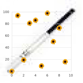
Sinemet 125mgNote the arrangement of nudel of the adenoma perpendicular to the basement membrane (polarity) symptoms chlamydia sinemet 300mg for sale. Endogenous occasions account for the inherited susceptibility to colon cancer, whereas exogenous occasions account fur the dietary and environmental contributors to colon most cancers. Dietary and environmental factors can also work together with inherited genetic modifications to promote the development of colon cancers. But these genes might promote the event of colon cancer in particular: individuals exposed to certain dietary or environmental threat components. Howevet unlike oncogenes, estrogen signaling is c:learly insufficient to provoke tumorigenesis, because the breasts ofall healthy girls are constantly beneath energetic estrogenic signaling without the event of tumors. Deregulation of this advanced biology results in a diverse group of neoplastic breast ailments inherently related with growth factor or hormonal signaling. These range from benign fibrocystic changes of the breast to malignant tumors of the breast. Three broadly defined molecular pathways appear to be related to the pathophysiology of breast cancers: hormone signaling. Growth Factor Slgnallng A quantity ofgrowth factor signaling pathways are implicated in the pathophysiology of breast cancers. In contrast, lowered publicity to estrogen and progesterone protects in opposition to the event ofbreast most cancers. The clinic:al success of anti-estrogen therapies provides proof of precept of the essential function of estrogen signaling within the pathogenesis of breast most cancers. Mutations in these genes are additionally associated with a excessive incidence of ovarian cancer in women and increased incidences of prostate cancer, melanomas, and breast cancer in men. Both of those genes perform as tumor suppressor genes such that breast tumors contain each the inherited abnormality in one allele and a somatic loss ofthe remaining allele. The Cellular Evolutlon of Breast most cancers Cancer of the breast is kind of always a result of the malignant transformation of secretory epithelial cells. The regular secretory breast epithelium consists of two layers: the basal myoepithelial layer and the luminal epithelial layer. These layers can be identified by their characteristic patterns of expression of keratin filaments. The identity of the cell of origin of breast cancers has been debated for decades and has not yet been completely outlined. Our current understanding suggests that all breast cancers come up from a cell subset within the luminal epithelial layer. However, in the center of tumorigenesis, some breast cancers can de-differentiate and ultimately develop attribute options of the basal layer cells. Abnormalities in the ductal epithelium are evident even earlier than the onset of hyperproliferative states that exhibit excessive numbers and layers oflurninal cells. Genetic abnormalities probably underlie the earliest form of ductal pathology, referred to as flat atypia, wherein the normal single-layered epithelium architecture is preserved, although the cell population is replaced by atypical cells. Breast most cancers Subtyplng by Morphology or Receptor Expression Breast cancers are extraordinarily heterogeneous of their morphology and biology. Such a diverse illness requires classification methods to higher understand and handle it. Additional genetic occasions lead to progressively Increased and irregular overgrowth (hyperplasla, then carcinoma In sltu) and eventually to Invasive ductal carcinoma. Changes In chromatin are evident In the nuclei early on and are one of the options of atypla. Three are proven here-hlstologlc subtypes (top left), intrinsic subtypes (top right), and dinical subtypes (bottom)-with their proportional incidences shown via pie chart. By their distinct morphologies, the majority of breast cancers can be categorized as ductal carcinomas. Ductal carcinomas exhibit various degrees of tubule formation, but lobular carcinomas have misplaced the ability to kind tubules. This is as a end result of of a failure in cell-cell attachment as a consequence of the mutational lack of the cell adhesion protein E-cadherin. Breast Cancer Subtyping by Transcriptomlc Analysis the event of methods to simultaneously determine the expression of 10,000 or extra genes has revolutionized the way cancers may be classified. These molecular subtypes have robust prognostic significance, with the luminal A subtype having the most effective prognosis and the basallike subtype the worst prognosis. The basal-like subtype lacks a unifying molecular attribute, but its hallmark is a major quantity ofgenomic instability. And since gene mutations persist in the respiratory epithelium for years after cessation of smoking, smokers remain at risk for lung most cancers for as a lot as twenty years after smoking cessation. At least four distinct kinds of cancer develop in the lung, arising from distinct cells oforigin in the bronchiolar and alveolar respiratory epithelium. Squamous-cell lung cancers are thought to arise from basal progenitor cells lining the bronchiolar epithelium. Small-cell lung cancers are thought to come up from pulmonary neuroendocrine cells discovered scattered in the bronchioles. Adenocarcinomas are thought to come up from alveolar kind 2 cells, which are thought to have progenitor functionality. Large-cell lung cancer is a collective time period applied to the remaining rarer subtypes of lung cancer. Consistent with the placement of their cells of origin, squamous-cell and small-cell lung cancers are regularly found in additional central areas of the lungs, whereas adenocarcinomas regularly arise within the distal areas of the lungs. Since small-cell lung cancers come up from a distinctly completely different (neuroendocrine) cell of origin and their biologic habits is also very different, the opposite three varieties are usually collectively referred to as non-small-cell lung cancers to clearly differentiate them from small-cell lung most cancers. In addition to its prognostic significance, this evaluation of breast cancers by molecular signatures has predictive worth regarding the sensitivity of every to varied anti-cancer treatments. Indeed, different predictive gene signatures have been developed as commercial assays for scientific breast cancer samples and might provide validated prognostic and predictive scores, enabling more personalized remedy planning for individual sufferers. This is as a result of the lung epitheliwn is highly uncovered to environmental carcinogens, particularly these in tobacco smoke. Exposure to these carcinogens induces gene mutations within the epithelium alongside the complete respiratory tract. The proxlmal airways are Involved In the passage of air, protection, and drainage of secretions, whereas the dlstal alveoll are Involved In fuel trade. According to our present understanding, the different types of lung cancer artse from different varieties of cells throughout the respiratory system. For instance, lung adenocarcinomas are frequently preceded by pre-neoplastic lesions known as atypical adenomatous hyperplasia; their cells frequently harbor the same oncogene mutations found within the later adenocarc:inomas. Numerous oncogenes which are disease drivers for subsets of adenocarcinomas and squamous-cell lung cancers have been recognized.
Generic sinemet 125mg on-lineEpldemlology Sjogren syndrome occurs in approximately 1-3% of the adult inhabitants treatment for depression buy generic sinemet from india. The prototypic affected individual is a lady in the fourth or fifth decade of lire. Pathophyslology Although the cause ofSjogren syndrome stays unclear, several pathways have been implicated in its pathogenesis. Central among these is autoimmunity to epithelial tissues, with an immwae response directed against a quantity of ubiquitously expressed antigens (eg, Fodrin, Ro, and La), as properly as to some antigens expressed. The antibodies to M3R are believed to forestall the stimulated secretion of saliva and tears and could also be important generators of the hyposecretion that characterizes the disease. Histologically, an intense mononuclear inflammatory infiltrate is noticed in affected lacrimal and salivary glands, respectively. Like different autoimmune rheumatic ailments, outstanding polyclonal hypergammaglobulinemia and high-tite. Cllnlcal Manifestations the most outstanding presenting symptoms in Sjogren syndrome are ocular and oral dryness. Intense xerophthalmia (ocular dryness) may express itself as eye irritation, with a international physique sensation or with pain. Such impairment in tear manufacturing heightens the chance ofcorneal ulcer and perforation. Impaired saliva production, at rest and with stimulation when consuming, contributes to the outstanding symptom of:urostomia (dry mouth). Affected individuals typically report difficulty in swallowing dry meals or in talking at size without entry to a beverage. Characteristically, people affected by Sjogren syndrome are susceptible to new-onset and severe dental caries at the gum line in mid-adult lire. Other epithelial surfaces may be similarly affected by diminished secretions and contribute to dryness. Dryness within the respiratory tract may give rise to hoarseness and reaurent bronchitis. Other doubtlessly affected organ systems embrace the kidneys, lungs, joints, and liver (resulting in interstitial nephritis, interstitial pneumonitis, nonerosive polyarthritis, and intrahepatic bile duct inflammation). Those sufferers with significantly severe disease are at elevated danger of cutaneous vasculitis (including palpable purpura and pores and skin ulceration) and lymphoproliferative disorders (eg, mucosaassociated ly. Maintaining oral hydration, with access to a regular supply of beverages, is inspired. For those affected by extreme illness sequelae (including systemic vasculitis and mononeuritis multiplex), the administration of systemic immunosuppression is necessary. While such inflammation predominantly involves striated muscle, you will need to recognize that clean muscle and even cardiac muscle may equally, although less commonly. Often, the stricken patient experiences increasing problem when rising from a seated position, getting off the bed, or ascending a flight of stairs. At essentially the most extreme end of the disease spectrum, affected individuals could develop profound impairment in swallowing stable foods and in full lung expansion, arising from the pathologi. These illness manifestations may outcome in the nasal regurgitation of swallowed liquids and in profound respiratory compromise with hypoventilation. There can additionally be a predilection for extramuscular involvement to occur, together with of the lung parenchyma (interstitial pulmonary fibrosis) and peripheral joints (inflammatory polyarthritis). A third type, referred to as immune-mediated necrotizing myopathy has now been recognized. Polymyositis has been estimated to happen with an annual incidence ofapproximately 5 instances per million. Interestingly, dermatomyositis has a bimodal distribution in terms of age at onset; the first peak occurs in childhood, and the second peak happens in mid- and late adult life. Autoantibodies are present in approximately 60% of all patients with an inflammatory myositis. Several of those antibodies are closely linked to distinct medical phenotypes, and their presence may present perception into both the diagnosis and prognosis of the illness in query. The attribute skin and nailfold capillary adjustments seen in patients with dermatomyositis lend support to this notion. Recent evidence has implicated the type 1 interferon pathway within the pathogenesis of dermatomyositis. A particular antibody affiliation has been described in up to 40% of patients with one form of necrotizing myopathy. These sufferers have a severe form of necrotizing myopathy which will persist even after the statin medication is withdrawn. Recent research counsel that one source of these autoantigens is the regenerating muscle cell itself, which expresses larger levels of myositis autoantigens than its regular counterpart. An intriguing pathophysiologic hypothesis is that the immune response that targets related antigens in both tumor and inflamed muscle cells could be liable for the hyperlink between inflammatory myositis and malignancy. Pathophysiology the inflammatory myopathies share a quantity of comparable pathologic features but also possess distinct ones. However, careful interpretation of biopsy specimens is critical since (1) several of those modifications (especially damage/regeneration) can be seen in the muscle of sufferers with noninflammatory "mimics" such because the muscular dystrophies; and (2) inflammatory infiltrates are only rarely seen in the necrotizing myopathies. It has been instructed that the inflammation seen in polymyositis is driven by autoantigens expressed within the muscle setting, given the restricted T-cell repertoire in both circulating and muscleinfiltrating lymphocytes. Further damage occurs when infiltrating T cells degranulate and release perforin and proteolytic granzymes at particular sites of contact inside the affected muscle. Major involvement of the capillaries has led many experts to recommend that the primary disorder in dermatomyositis is a small-vessel Clinical Manifestations the inflammatory myopathies characteristically begin over a quantity of weeks to a couple of months. This characteristically involves the upper and lower extremities and is predominantly proximal quite than distal in location. Routine every day actions that one would possibly otherwise take without any consideration can turn out to be quite a chore, and even an unimaginable ordeal, to carry out. In addition, the cutaneous options of dermatomyositis could be fairly debilitating and include a painful, burning sensation of the affected pores and skin, as well as skin cracking and even breakdown with open ulceration. In patients with dermatomyositis, a fifth criterion is a characteristic skin rash. Erythematous and/or violaceous discoloration may happen around the exterior of the attention, or in a V-neck distribution on the trunk. These prototypic pores and skin adjustments are termed the periorbital heliotrope and the scarf sign, respectively. Extensive sheets of muscle and delicate tissue calcification could occur in youngsters with dermatomyositis. Though latest efforts to modify the unique diagnostic standards, by integrating newer imaging modalities together with magnetic resonance imaging or utilizing newer autoantibodies with specificities for the inflammatory myopathies, have been proposed, the unique standards remain the inspiration for these two muscle disorders. In grownup patients, the model new analysis ofan inflammatory myopathy regularly heralds the co-oc:currence or subsequent development inside 1-5 years of a malignancy. The veracity of this remark has been confirmed in a number of population-based research that hyperlink the diagnoses of dermatomyositis and polymyositis with most cancers in cancer registries. A prognosis of dermatomyositis carries a twofold greater risk of incident malignancy.
Syndromes - Primary amyloidosis
- Thyroid function
- Discomfort during the test
- Family history (age-related hearing loss tends to run in families)
- Arrive on time at the hospital.
- Irritability
- The dislodged fat is vacuumed away through the suction tube. A vacuum pump or a large syringe provides the suction action.
- Infectious disease -- infections affecting the tissues of any part of the body
- Spinal anesthesia. This is also called regional anesthesia. The painkilling medicine is injected into a space in your spine. You will be awake but will not be able to feel anything below your waist.
Order sinemet 110 mgType I autoimmune pancreatitis accounts for more than 80% of circumstances in the United States and is associated with elevated serum levels of IgG4 and with lymphocytic infiltration throughout the pancreatic parenchyma treatment zone guiseley 300mg sinemet free shipping. Many patients with sort I autoimmune pancreatitis have extrapancreatic manifestations and are sometimes categorised as having IgG4-related disease. Of note, sort I autoimmune pancreatitis most commonly presents with distal biliary obstruction and jaundice, mimicking periampullary malignancy. The pathognomonic histopathologic findings in this illness are granulocyte-epithelial lesions with neutrophilic infiltration. In about 15-25% of instances of acute pancreatitis, no etiologic issue can be recognized. Idiopathic acute recurrent pancre-atitis is seen in patients with a couple of attack of acute pancreatitis when the underlying trigger eludes detection regardless of a thorough search. However, each the diploma of harm and the scientific penalties are quite variable. When the harm is limited in extent, the pathologic options consist of gentle to marked gland swelling, particularly within the acini, and gentle to marked infiltration with polym. In some cases, suppuration may be found together with edema, and this will lead to tissue neaosis and abscess formation. In severe instances, massive neaosis and liquefaction of the pancreas occur, predisposing to pancreatic abscess formation. Vascular necrosis and disruption may occur, resulting in peripancreatic hemorrhage. While microvascular hemorrhage involving peripancreatic tissue is frequent in severe circumstances of acute pancreatitis, significant bleeding from large-vessel erosion is a rare scientific entity and is more usually seen in chronic pancreatitis. Severe cases of pancreatitis may be associated with the formation of ascltes, which is in all probability going a mix of serous fluid excreted by the infected peritoneal floor, liquefied peripancreatic fat, blood from peripancreatic tissues, and necrotic pancreatic debris. In uncommon cases related to duct:al disruption of the pancreas, the ascites may contain frank. Documentation of amylase-rich peritoneal fluid establishes the diagnosis of so-called pancreatic asdtes. In instances of extreme acute pancreatitis, the peritoneal surfaces have a attribute look upon surgical exploration or post-mortem; fats necrosis, or saponification, might happen in and around the pancreas, omentum, and mesentery, showing as chalky white foci that will later calclfy. Histologic studies of pancreas tissue obtained from patients with first attacks of acute alcoholic: pancreatitis who underwent surgical procedure for problems have found that the acute pancreatitis (pancreatic: necrosis, steatonec:rosis, infiltration by inflammatory c:ells) sometimes develops in a gland already affected by chronic pancreatitis (perilobular and intralobular fibrosis, loss of exocrine parenchyma and atrophy of residual lobules, dilated interlobular and intralobular ducts lined with cuboidal or flattened epitheliwn, and protein plugs within dilated ducts). The calcium thus released leads caeruleln to the pathologlc results Inducing pancreatitis. However, elegant research have confirmed that the expression of energetic trypsin inside pancreatic acini is itself enough to induce cell dying and irritation in acute pancreatitis. Thus, the in vivo role of different mechanisms of pancreatic autodigestion remains unclear. Calc:ineurin is a probable downstream target of elevated intracellular Ca2� levels, mediating a few of the harm observed in ac:ute pancreatitis through T-cell activation. Trypsinogen is likely activated inside membrane-bound intracellular compartments that ei:hlbit dysregulated autophagy within the setting of acute pancreatitis. The formation oflysolecithin from the lecithin in bile might contribute to the disruption of the pancreas and the necrosis of surrounding fat. Phospholipase A2 also liberates arachidonic acid, which is then converted to prostaglandins, Ieukotrienes, and different mediators of inflammation, contributing to coagulation necrosis. Pancreatic lipase, launched as a direct result of pancreatic acinar cell harm, acts enzymatically on surrounding adipose tissue, causing the characteristic peripancreatic fats necrosis seen in extreme acute pancreatiti. Furthermore, trypsin and chymotrypsin activate kinins, complement, coagulation elements, and plasmin, resulting in edema, inflammation, thrombosis, andhemorrhage inside the gland. Circulating phospholipases intervene with the traditional perform of pulmonary surfactant, contributing to the development of an adult respiratory distress syndrome in some sufferers with acute panc. Elevated serum lipase levels are sometimes related to fats necrosis exterior the abdomen. C households of cytokines are implicated within the pathogenesis of the native and systemic inflammatory response. Cytoldnes this probably occurs solely in certain pathologic conditions, similar to a low intracellular pH. The mechanism of pH disturbance inside acinar cells is likely due to an alteration in cell signaling mechanisms and an inhibition ofacinar bicarbonate secretion. Moreover, although cathepsin L (an various isoform ofcathepsin B) normally degrades trypsin in an important cellular protective mechanism, a disturbance within the intracellular environment has been proven to contribute to an imbalance in cathepsin B activity relative to cathepsin L activity. The pathogenesis of alcoholic pancreatitis may be unique and should contain disordered agonist-receptor interaction on the membrane of pancreatic acinar cells. Whether ethanol or other alcohols mediate these results by interfering with acinar cell signaling pathways or by affecting acinar cell membrane fluidity is presently beneath investigation. The pathologic modifications outcome from the action of activated trypsin and other pancreatic eneymes on the pancreas and surrounding tissues. Similarly, elastase, once activated from proelastase, digests the elastin in blood vessel walls and causes vascular damage and hemorrhage; harm to peripancreatic blood vessels can lead to hemorrhagic pancreatitis. This launch appears to be in response to the presence of energetic digestive enzymes, unbiased of the underlying trigger. The manufacturing of cytokines throughout clinical pancreatitis begins shortly after ache onset and peaks 36-48 hours later. Systemic issues of acute pancreatitis, corresponding to respiratory failure, shock, and even multi. Substance P appears to be a strong pro-inflammatory mediator of both pancreatitis and related lung harm. On the opposite hand, complement issue 5a (C5a) appears to act as an antiinflammatory agent through the improvement of pancreatiti. Various factors play energetic roles as pro-inflammatory or anti-inflammatory brokers in acute pancreatitis. Clinical Manifestations Acute pancreatitis could present in a highly variable manner, with the severity of irritation and related morbidity differing markedly among sufferers. The distinction between acute pancreatitis and an acute exacerbation of chronic pancreatitis is set by the clinical history and the characteristic findings of chronic pancreatitis on imaging. In apply, the first two parts are sometimes current and enough to make to the clinical diagnosis. Each of the mediators proven plays a role In the event of the systemic manifestations of acute pancreatltls. The x-ray film shows a sharply demarcated space of localized colonic dilation and edema ("colon cutoff signal"). However, persistent fever beyond the fourth or fifth day ofillness-or spiking temperatures to 40�C or more-may signify the development of infectious problems suc. The cardinal laboratory discovering in acute pancreatitis is an elevation of the serum. The sensitivity of the serum amylase in acute pancreatitis is estimated to be 70-95%, meaning that 5-30% of sufferers with acute pancreatitis have normal or minimally elevated serum amylase values. Patients with marked greater than 3-fold) elevations of serum amylase usually have acute pancreatitis.
Buy discount sinemet 300mg lineLiver disease can be acute or cluonic medicine mart proven 110 mg sinemet, focal or diffuse, delicate or extreme, and reversible or irreversible. Most circumstances ofacute liver illness (eg, brought on by viral hepatitis) are so mild that they never come to medical consideration. In other instances ofacute liver harm, signs and signs are extreme enough to call for medical consideration. This syndrome of acute liver failure (also referred to as fulminant hepatic failure) carries a high mortality fee; nevertheless, lately. Liver harm may continue past the preliminary acute episode or could additionally be recurrent (chronic hepatitis). In some cases of persistent hepatitis, liver perform remains steady or the disease course of finally resolves altogether. In cirrhosis, the liver becomes onerous, shrunken, and nodular and displays impaired operate and diminished reserve due to a decreased quantity of functioning liver tissue. More importantly, the physics of blood move is altered such that the strain within the portal vein is elevated. As a end result, the blood is diverted around the liver rather than filtered via the liver. Although liver illness resulting from many alternative causes could current in widespread ways, the reverse can be true (le, liver illness from particular causes may have distinctly completely different displays in numerous patients). Such variations in the severity ofliver disease are most likely as a result of genetic, immunologic, and environmental (including perhaps nutritional) components which are currently poorly understood. Like many organs of the body, the liver normally has both a huge reserve capability for the varied biochemical reactions it carries out and the ability to regenerate totally differentiated cells and thereby recuperate completely from acute injury. Thus, only in essentially the most fulminant instances or in endstage illness are there insufficient residual hepatocytes to preserve minimal important liver features. More commonly, sufferers show transient indicators ofliver cell necrosis and disordered perform followed by full recovery. The signs and indicators of this kind of acute liver damage can best be understood as an impainnent of the conventional biochemical features of the liver. Other penalties ofliver illness are irreversible, usually seen in the affected person with cirrhosis. Primary blllary cirrhosis Autoimmune hepatitis Scleroslng cholangltls Overtap syndromes Graft-versus-host disea5e Allograft rejection Genetic llVllrd. Hepatocellular patterns (lsonlazld, acetaminophen) Cholestatic patterns (methyltestosterone) Mixed patterns (sulfonamides, phenytoin) Mlcroveslcular and macroveslcular steatosls (methotrexate) Vucularlnjury Venooccluslve illness Budd-Chlarl syndrome lschemlc hepatitis Passive congesUon Portal vein thrombosis Nodular regenerative hyperplasia ��leslons Hepatocellular carcinoma Cholanglocardnoma Adenoma Focal nodular hyperplasia Metastatic turnors Abscess Cysts Hemanglorna Repioduced, with permtulon, from Ghany Met al. Commonly, sufferers with cirrhosis present with superimposed acute liver harm (eg, caused by an alcoholic binge or different drug exposure). What elements might determine the difference in severity of liver illness between two patients with acute hepatitis ensuing from the identical cause In what methods Is the affected person with underlying cirrhosis who presents with acute hepatitis likely to be differ- ent from the patient with a prevlously regular liver and acute hepatitis The right lobe has two lesser segments: the posterior caudate lobe and the inferior quadrate lobe. The liver may additionally be differentiated functionally via the portal blood circulate into 4 sectors, which are additional subdivided into eight segments. The liver weighs approximately 1400 g in the adult and is roofed by a fibrous capsule. These vessels converge within the liver, and the combined blood move exits through the so-called central ftim (also referred to as terminal veins or hepatic venules) that drain into the hepatic vein and ultimately the inferior vena cava. The portal vein carries venous blood from the small intestine, rich in freshly absorbed nutrients-as properly as medication and poisons-directly to the liver. Also:flowing into the portal vein earlier than its entry into the liver is the pancreatic venous drainage, rich in pancreatic hormones (insulin, glucagon, somatostatin, and pancreatic polypeptide). The portal vein varieties a specialized capillary mattress that permits individual hepatocytes to be bathed directly in portal blood. It is in these sinusoids that blood from the hepatic artery is mixed with blood from the portal vein on the way in which to the central vein. The ret:iculoendothelial cell meshwork in which the hepatocytes reside consists of various cell sorts, most importantly the endothelial cells that make up the partitions of the sinusoids; specialised macrophages, termed Kupffer cells, which are anchored in the sinusoidal space; and stellate cells or lipocytes, fat-storing cells involved in vitamin A metabolism, which lie between the hepatocytes and the endothelial cells. Approximately 30% of all cells in the liver are reticuloendotheUal cells, and about 33% of those are Kupffer cells. Yet, as a end result of reticuloendothelial cells are smaller than hepatocytes, the reticuloendotheli. They carry out specific functions, including phagocytosis and cytokine secretion, and talk with each other in addition to with hepatocytes. Their dysfunction contributes each to hepatocyte necrosis in acute liver disease and to hepatic fibrosis in continual liver illness. This could additionally be seen in liver biopsies from patients with liver illness of unknown trigger. The final hepatocytes reached by the blood earlier than it enters the central vein are termed zone 3 hepatocytes. Thus, the microscopic: group of the liver could be viewed by means of useful zones. Hepatocytes at either extreme of the acinus (zones 1 and 3) appear to differ in each enzymatic exercise and physiologic functions. Zone 1 hepatocytes, exposed to the highest oxygen concentrations, are particularly lively in gluc:oneogenes. They are also the major web site of urea synthesis (because freely diffusible substances similar to ammonia absorbed from protein breakdown within the intestine are largely extracted in zone 1). Conversely, zone three hepatocytes are extra lively in glycolysis and lipogenesis (processes requiring less oxygen). These substances enter whichever hepatocytes have the suitable transporters no matter their zone. Hepatocytes: Polarized Cells with Functional Segregation All surfaces ofa hepatocyte are notthe similar. Very different activities go forward at these areas of the hepatocyte plasma membrane; tight junctions between hepatocytes serve to preserve segregation of apical and basolateral plasma membrane domains. Receptor-Mediated Uptake Functional zonation applies only to processes driven by the presence of diffusible substances. There is, nevertheless, no clear line between the implications of disturbed apical and basolateral features: Cholestasis, although initially a dysfunction of apical bile circulate, is ultimately manifested at the basolateral surface. Similarly, disruption of energy metabolism or protein synthesis, although initially impinging on the secretory and metabolic processes of the hepatocyte, will ultimately have an effect on the bile transport machinery in the apical plasma membrane as well. Accordingly, there must be little resistance to its circulate inside the liver, allowing the blood to percolate through the sinusoids and achieve maximal contact-for change of substances-with hepatocytes. Two distinctive features-fenestrations within the endothelial cells and lack ofa typical basement membrane between endothelial cells and hepatocytes-aid in making the liver a low-pressure circuit for the flow of portal blood. These options are altered in cirrhosis, resulting in increased portal stress and profound adjustments in liver blood flow, with devastating medical consequences.
Discount 300 mg sinemet amexPatients normally study to minimize orthopnea by sleeping with the higher body propped up by two or more pillows symptoms 2015 flu cheap sinemet. Sudden onset of severe respiratory distress at night-paroxysmal nocturnal dyspnea-probably happens because of the reduced adrenergic help of ventricular perform that occurs with sleep, the rise in blood return as described previously, and normal nocturnal depression of the respiratory heart. Confusion may come up in advanced heart failure due to under-perfusion of the cerebrum. Nocturla-Heart failure can lead to reduced renal perfu- 305 sion in the course of the day whereas the affected person is upright, which normalizes solely at evening whereas the patient is supine, with consequent diuresis. Cheat pain-Ifthe cause offailure is coronary artery disease, sufferers may have chest pain secondary to ischemia (angina pectoris). In addition, even with out ischemia, acute heart failure could cause chest pain by unknown mechanisms. Kales, pleural effusion-Increased fluid in the alveolar spaces from the mechanisms described beforehand can be heard as ral. Increased capillary pressures can also trigger fluid accumulation within the pleural areas. Dilpluecl and sustained apical impulle-In most people, contraction of the center may be appreciated by cautious palpation of the chest wall (apical impulse). The regular apical impulse is felt within the midclavicular line within the fourth or fifth intercostal house and is palpable solely during the first part of systole. Sustained impulses recommend that will increase in left ventricular quantity or mass are current. In addition, when left ventricular volume is elevated as a compensatory mechanism of heart failure, the apical impulse is displaced laterally. The exact mechanism answerable for the genesis of the third heart sound is unknown, however the sound appears to end result either from the sudden deceleration of blood because the elastic limits of the ventricular chamber are reached or from the actual impact of the ventricular wall against the chest wall Although a 3rd coronary heart sound i. In these people, the presence of a third coronary heart sound is almost pathognomonic of ventricular failure. The increased end-systolic volumes and pressures characteristic of the failing heart are most likely answerable for the distinguished third heart sound. When it arises as a result of ofleft ventricular failure, the third coronary heart sound is usually heard greatest at the apex. As with the third heart sound, the precise mechanism for the genesis of the fourth heart sound is unknown. However, it most likely arises from the sudden deceleration of blood in a noncompliant ventrlde or from the sudden impact of a stiff ventricle against the chest wall It is greatest heard laterally over the apex on the point ofmaJrimal impulse, notably when the patient is partially rolled over onto the left facet. The fourth heart sound is usually heard in any affected person with coronary heart failure resulting from diastolic dysfunction. Pale, chilly, and sweaty skin-Patients with severe coronary heart failure typically have peripheral vasoconstriction, which maintains blood move to the central organs and head. In some cases, the skin appears dusky because of lowered oxygen content in venous blood because of increased oxygen extraction from peripheral tissues that are receiving low blood flow. The interventricular septum is normally bowed toward the thinner-walled and lower-pressure proper ventricle. When right ventricular pressure increases relative to the left, the interventricular septum can bow to the left and stop environment friendly filling of the left ventricle, which can lead to pulmonary congestion. Rarely, the bowing may be so severe that left ventricular outflow is partially obstructed. Right Ventricular Failure ainical Presentation Symptoms of right ventricular failure embody shortness of breath, pedal edema, and abdominal ache. Patients with right ventricular failure could have a third coronary heart sound heard finest on the sternal border or a sustained systolic heave of the sternum. Beause the most common reason for proper ventricular failure is left ventricular failure, indicators of left ventricular failure are sometimes additionally current. Shonness of Breath-In left ventricular failure, sufferers ma:y be in want of breath due to pulmonary edema, as discussed previously. In patients with right-sided failure resulting from pulmonary disease, shortness of breath could also be a manifestation of the underlying illness (eg. In some patients with proper ventricular failure, congestion of the hepatic veins with formation of ascites can impinge on normal diaphragmatic function and contribute to the sensation of dyspnea. In addition, decreased right-sided cardiac output alone may cause acidosis, hypoxia, and air hunger. If the trigger of right-sided failure is a left-sided defect such as mitral stenosis, the onset of proper coronary heart failure can sometimes lessen the signs of pulmonary edema because of the decreased load positioned on the left ventricle. As mentioned, left ventricular failure may cause right ventricular failure beause of the increased afterload positioned on the best ventricle. Increased afterload can additionally be current from abnormalities of the pulmonary arteries or capillaries. For instance, elevated circulate from a congenital shunt could cause reactive pulmonary artery constriction, elevated right ventricular afte. Right ventricular failure can occur as a sequela of pulmonary illness (cor pulmonale) as a result of ofdestruction of the pulmonary capillary bed or hypoxia-induced vasoconstriction of the pulmonary arterioles. Pathophysiology the pathophpiology of right ventricular failure is much like that described for the left ventricle. Both systolic and diastolic abnormalities of the right ventricle could be present and normally occur beause of inappropriate masses placed on the ventricle or main lack of myocyte contractility. The vertical distance above the center at which venous pulsations are observed is an estimate of the best atrial or central venous stress. Right atrial strain can then be approximated by adding 5 cm to the peak of the venous column (because the best atrium is approximately 5 cm inferior to the angle). Jugular venous pulsations are often observed lower than 7 cm above the best atrium. Elevated atrial pressures are current any time this distance is bigger than 10 cm. Elevated atrial pressures point out that the preload of the ventricle is sufficient but ventricular operate is decreased and fluid is accumulating in the venous system. Other causes of elevated jugular pressures in addition to heart failure embody pericardia! In addition to relative position, individual waveforms of the jugular venous pulse could be assessed. The a wave is caused by transmitted proper atrial stress from atrial contraction. What are the main medical manifestations and complications of left-versus right-sided coronary heart failure In this part, the pathophysiologic mechanisms of stenotic and regurgitant aortic and mitral valves are addressed.
Discount sinemet 300mg onlineAs a result treatment atrial fibrillation cheap sinemet 300mg amex, air flow of respiratory items becomes nonuniform, and the matching of air flow to perfusion is altered. Pure shunt is uncommon in bronchial asthma despite the very fact that mucus plugging is a common finding, particularly in severe, deadly bronchial asthma. Arterial C01 rigidity is usually regular to low, given the increased air flow seen with bronchial asthma exacerbations. Even delicate hypercapnia ought to be viewed as an ominous signal during a severe bronchial asthma attack, indicating progressive airway obstruction, muscle fatigue, and falling alveolar ventilation. Clinical Manifestations the manifestations of bronchial asthma are readily defined by the presence of airway irritation and obstruction. Symptoms & Signs-The variability of symptoms and signs is an indication of the tremendous range of disease severity, from gentle and intermittent disease to chronic, extreme, and sometimes deadly asthma. The higher muscular effort required to overcome increased airway resistance is detected by spindle stretch receptors, principally of intercostal muscles and the chest wall. Lung compliance falls, and the work of respiratory increases, additionally detected by chest wall sensory nerves and manifested as chest tightness and dyspnea. Rising arterial col rigidity and, later, evolving arterial hypoxemia (each alone or collectively as synergistic stimuli) will stimulate respiratory drive through peripheral and central chemoreceptors. Wheezing-Smooth muscle contraction, along with mucus hypersecretion and retention, leads to airway caliber reduction and prolonged turbulent airflow, producing auscultatory and audible wheezing. Cough-Cough outcomes from the combination of airway narrowing, mucus hypersecretion, and the neural afferent hyperresponsiveness seen with airway irritation. Such testing reveals nonspecific hyperresponsiveness in virtually all individuals with bronchial asthma, including these with mild disease and normal spirometry findings. Other brokers which were used to establish particular publicity sensitivities embody su1fur dioxide and toluene diisocyanate. What are the pathologic events that contribute to chronically irregular airway architecture In asthma However, it is essential to recognize that each processes could be found within the identical particular person, with a varying contribution of each to the overall clinical phenotype. Chronic Bronc:hltll-Chronic bronchitis is defined by a medical historical past of productive cough for three months of the 12 months for two consecutive years. Both dyspnea and airway obstruction, typically with an element of reversibility, are intermittently to repeatedly present. The main web site ofinflammatory involvement determines the predominant pathophysiologic process in an individual, with airway-predominant disease causing cluonic bronchitis and pare. The airway mucosa is variably infiltrated with inflammatory cells, including polymorphonuclear leukocytes and lymphocytes. In the absence of regular ciliated bronchial epithelium, mucodliary clearance operate is severely diminished or utterly abolished. Hypertrophy and hyperplasia of submucosal glands are distinguished features, with the glands typically making up more than 50% of the bronchial wall thickness. Bronchial smooth muscle hypertrophy is common, and hyperresponsiveness to nonspecific bronchoconstri. In the absence of any superimposed course of, corresponding to pneumonia, the gas-exchanging lung parenchyma. Emphysema-Pulmonary emphysema is a situation marked by irreversible enlargement of the airspaces distal to the terminal bronchioles. Pathologic and etiologic distinctions may be made among varied patterns of emphysema, however the clinical displays of all are similar. Furthennore, by 2030 the prevalence of the disease is projected to rise to an anticipated four. It is estimated to have direct prices ofapproximately $32 billion dollars per 12 months. Mortality additionally increases with age and is greater in individuals oflower socloeconomic standing. Cigarette smoking is the most common danger factor worldwide, representing the principal cause in as much as 9096 of sufferers. Reduced circulating and tissue levels can lead to the early onset of severe emphysema. Alpha, -protease inhibitor is able to inhibiting a quantity of forms of proteases, including neutrophil elastase, which is implicated within the genesis ofemphysema (see Pathophy5iology section below). Autosornal dominant mutations, especially in northern Europeans, produce abnormally low serum and tissue levels of this inhibitor, altering the balance ofconnective tissue synthesis and proteolysis. In persistent bronchltfs, the thickness of the mucous glands Increases and could be expressed as the ratfo of (b-c/(a-d); this Is known as the Reid Index. The results of these combined modifications is continual airway obstruction and impaired clearance of airway secretions. The nonuniform airway obstruction of persistent bronchitis has substantial results on air flow and gasoline exchange. Ventilation/perfusion relationships are altered with increased areas of low V/Q ratios. These low V/Q mismatches are largely responsible for the more important resting hypoxemia seen in continual bronchitis, compared with that seen in emphysema. Oxidants, whether or not endogenous (supero:xide anion) or exogenous (eg, cigarette smoke). In contrast to chronic bronchitis, a disease of the airways, emphysema is a disease of the encompassing lung parenchyma. The physiologic consequences result from three necessary modifications: (1) destruction of terminal respiratory units; (2) lack of alveolar-capillary bed; and (3) lack of the supporting constructions of the lung, including elastin-containing connective tissue. This loss of connective tissue reduces the traditional help of noncartilaginous airways, resulting in a lung with diminished elastic recoil and elevated compliance. A premature expiratory collapse of airways ensues, with characteristic obstructive signs and physiologic findings. The pathologic picture of emphysema is considered one of progressive destruction of terminal respiratory models or lung parenchyma distal to terminal bronchioles. The interstitium of respiratory units harbors some inflammatory cells, however the chief discovering is a loss of alveolar walls and enlargement of airspaces. Alveolar capillaries are additionally lost, which can lead to decreased diffusing capacity and progressive hypoxemia, particularly with train. In centriacinar emphysema, destruction is focused within the heart of the terminal respiratory unit, with the respiratory bronchioles and alveolar ducts relatively spared. Panacinar emphysema includes destruction of the terminal respiratory unit globally, with diffuse airspace distention. This sample is usually, though not uniquely, seen in <Xi-protease inhibitor deficiency. Bullae are massive confluent airspaces shaped by higher native destruction or progressive distention of lung units.
Brown Algae (Laminaria). Sinemet. - Are there any interactions with medications?
- Preparation ("ripening") of the cervix in women, such as during childbirth or procedures.
- Are there safety concerns?
- How does Laminaria work?
- Dosing considerations for Laminaria.
- Weight loss, high blood pressure, cancer prevention, heartburn, and other conditions.
Source: http://www.rxlist.com/script/main/art.asp?articlekey=96544
Buy 125 mg sinemet with mastercardChronic Viral Hepatitis Varal hepatitis is the most common cause of persistent liver illness in the United States medications hyponatremia cheap sinemet 110mg otc. Alcoholic Chronic Hepatitis Chronic liver disease in response to some poisons or toxins might characterize the triggering of an underlying genetic predisposition to immune attack on the liver. In alcoholic hepatitis, nonetheless, repeated episodes of acute harm in the end trigger necrosis. Cellular and matrix alteratlons In the house of Dlsse are important events In the pathogenesis of hepatic fibrosis. The activation of llpoc:ytes, characterized by proliferation and Increased flbrogenesls, Is associated with the replacement of the traditional low-density matrix with a high-density matrix. These alterations are llkely to underlie, no much less than In part, the loss of each endothellal fenestrations (pores) and hepatocyUc mlcrovllll typical ofchronic llver Injury. Lipid accumulation has been shown to result in toxicity by diverse mechanisms, including increased oxidative stress because of the generation of reactive oxygen species by mitochondrial and peroxisomal fatty acid oxidation. An Increased uptake and/or decreased disposal of llplds (fatty acids) In the liver outcomes In 1n 1cc. Autoimmune hepatitis is a continual inflammatory liver disease characterized by a primarily T cell-mediated immune response to as yet unidentified auto-antigen(s). Most patients with autoimmune hepatitis show histologic enchancment in liver biopsies after treatment with systemic corticosteroids. The pcnistent activation of innate and adaptive immune responses mediates irritation and bile duct epithelial damage. The resultant bile duct destruction leads to continual cholestasis and the intrahepatic accumulation of cytotoxic bile acids. This accumulation causes liver cell injury and a secretion of pro-inflammatory cytokines that induce inflammation and fibrosis in surrounding tissue. While immune dyuegulation is implicated in its pathogenesis, major sclerosing cholangitis l. Owing to its definite affiliation with inflammatory bowel illness, a *leaky intestine" hypothesis has been postulated, whereby translocation of gastrointestinal flora results in disruption of biliary epithelial cells and exposes cholangiocytes to bile acid toxicity. Cholangiography displaying characteristic bile duct stricturing establishes the analysis of major scleroaing cholangitis. Pathology All forms of continual hepatitis ahare the widespread histopathologic features of (1) inflammatory infiltration of hepatic portal areas with mononuclear cells, especially lymphocytes and plasma cells; and (2) necrosia ofhepatocytes within the parenchyma or immediatdy adjacent to portal areu (periportal hepatitis, or "piecemeal necrosis"). A *cobblestone" look of liver cdls is seen, indicating hepatocyte regeneration. In extra severe circumstances of persistent hepatitis, the portal areu are expanded and densely infiltrated by lymphocytes, histiocytes, and plasma cella. More extreme circumstances additionally present evidence of necrosis and fibrosis between portal triads. The lymphocytes lengthen Into the peripheral a part of the lobule by way of the llmltlng plate. These connective tissue bridges are evidence of hepatic architecture remodeling, an important step in the improvement of cirrhosis. Fibrosis could extend from the portal areas into the lobules, isolating hepatocytes into clusters and enveloping bile ducts. Hepatocyte regeneration is seen with mitotic figures, multinucleated cells, rosette formation. Progression to cirrhosis is signaled by intensive fibrosis, lack of zonal architecture, and regenerating nodules. As with other shows of liver illness, not all sufferers with cirrhosis develop life-threatening complications. Clinical Manifestations Some sufferers with delicate continual hepatitis are totally asymptomatic and identified solely in the course of routine blood testing; others have an insidious onset of nonspecific symptoms such as anorexia, malaise, and fatigue or hepatic signs, corresponding to right upper quadrant stomach discomfort or ache. Fatigue in chronic hepatitis could additionally be related to a change in the hypothalamic-adrenal neuroendocrine axis led to by altered endogenous opioidergic neurotransmission. Other extrahepatic manifestations are unusual By definition, signs ofcirrhosis and portal hypertension (eg, ascites, collateral circulation, and encephalopathy) are absent. Laboratory studies show gentle to average will increase in serum aminotransferase, bilirubin, and globulin levels. The scientific manifestations of continual hepatitis probably replicate the position of a systemic genetically managed immune disorder within the pathogenesis of severe disease. Acne, hirsutism, and amenorrhea could occur as a mirrored image of the hormonal results of chronic liver disease. Laboratory research in sufferers with extreme chronic hepatitis are invariably abnormal to numerous degrees. Thus, the serum bilirubin, alkaline phosphatase, and globulin ranges may be regular and aminotransferase ranges only mildly elevated on the identical time that a liver biopsy reveals extreme chronic hepatitis. The pure historical past and treatment ofchronic hepatitis varies depending on its trigger. The complications of severe persistent hepatitis are those of progression to cirrhosis: variceal bleeding, encephalopathy, coagulopathy, hypersplenism, and ascites. These are largely because of portosystemic shunting quite than diminished hepatocyte reserve (see later discussion). Other causes include chronic biliary obstruction, medication, genetic and metabolic disorders, continual coronary heart failure, and first (auto. Pathogenesis the elevated or altered synthesis of collagen and different connective tissue or basement membrane parts of the extracellular matrix is implicated within the improvement of hepatic fibrosis and thus within the pathogenesis of cirrhosis. Thus, fibrosis may affect not solely the mechanics of blood flow by way of the liver but additionally the capabilities of the cells themselves. Hepatic fibrosis happens in three situations: (1) secondary to irritation and the following activation of immune responses; (2) as part of the process of wound healing; and (3) in response to agents that induce major fibrogenesi. Agents corresponding to carbon tetrachloride that attack and kill hepatocytes directly can produce fibrosis as a half of wound healing. In both immune responses and wound healing, the fibrosis is triggered not directly by the effects of cytokines released from invading inflammatory cells. Finally, sure brokers corresponding to ethanol and iron might cause primary fibrogenesis by directly growing collagen gene transcription and thus additionally growing the quantity of connective tissue secreted by cells. The actual culprit in all these mechanisms of elevated:fibrogenesis will be the fat-storing cells (stellate cells) of the hepatic reticuloendothelial system. What are the classes of continual hepatitis based on histologic findings on liver biopsy In addition to the stellate cells, fibrogenic cells also derive from portal fibrohluts, circulating fibrocytes, bone marrow, and epithelial-mesenchymal cell transition. The second stage involves the formation of subendothelial collagen cross-links, the proliferation of myoepithelial cells, and the distortion of hepatic architecture with the looks of regenerating nodules. Cirrhosis remains a dynamic state by which sure interventions, even at these advanced phases, might yield benefits similar to regression of scar tissue and improvements in medical outcomes.
110mg sinemet visaBoth chemokine manufacturing and adhesion molecule expression are upregulated by soluble inflammatory mediators 2 medications that help control bleeding buy sinemet visa. The scientific presentation of allergic rhinitis includes nasal, ocular, and palatal pruritus, paroxysmal sneezing, rhinorrhea, and nasal congestion. A private or household historical past of other allergic diseases similar to bronchial asthma or atopic dermatitis supports a diagnosis of allergy. Confirmation of allergic rhinitis requires the demonstration of particular lgE antibodies to frequent allergens by in vitro immunoassay or in vivo (skin) testing in sufferers with a historical past of signs with related exposures. Pathology & Pathogenesis Inflammatory adjustments within the airways are recognized as crucial options of both allergic rhinitis and persistent bronchial asthma. Crosslinking of surface-bound lgE by antigen activates tissue mast cells and basophils, inducing the immediate release of preformed mediators and the synthesis ofnewly generated mediators. Mast cells and basophils also have the flexibility to synthesize and launch proinflammatory cytokines, growth and regulatory components that interact in complicated networks. After intranasal problem or ambient publicity to a related allergen, the allergic affected person begins sneezing and develops a rise in nasal secretions. After roughly 5 minutes, the patient develops mucosal swelling leading to decreased airflow. Histologically, the early response is characterised by vascular permeability, vasodilatation, tissue edema, and a mild cellular infiltrate of largely granulocytes. The late-phase allergic response may observe the earlyphase response (dual response) or could occur as an isolated event. Late-phase reactions begin 2-4 hours after preliminary exposure to antigen, reach maximal activity at 6-12 hours, and normally resolve within 12-24 hours. If the exposure is frequent or ongoing, nonetheless, the inflammatory response turns into chronic. The late-phase response is characterized by erythema, induration, warmth, burning, and itching and microscopically by a big mobile inflow of mainly eosinophils and mononuclear cells. Changes consistent with airway remodeling and tissue hyperreactivity may occur. Clinical Manifestations the medical manifestations of allergic airway illness (Table 3-3) arise from the interplay of mast cell and basophil mediators with target organs ofthe upper and decrease airway. The symptoms of allergic rhinitis appear immediately after publicity to a related allergen (early-phase response), though many sufferers experience persistent and recurrent signs on the premise of the late-phase inflammatory response. Complications of severe or untreated allergic rhinitis embody sinusitis, auditory tube dysfunction, hyposmia, sleep disturbances, asthma exacerbations, and persistent mouth respiratory. Sneezing, Pruritus, and Mucus Hypersecretion Patients with allergic rhinitis develop persistent or episodic paroxysmal sneezing; nasal, ocular, or palatal pruritus; and watery rhinorrhea triggered by publicity to a selected allergen. Children frequently show indicators of obligate mouth respiratory, including long facies, slender ma:rillae, flattened malar eminences, marked overbite, and high-arched palates (so-called adenoid facies). Oral sympathomimetics that induce vasoconstrlction by stimulating a-adrenergic receptors are sometimes used in conjunction with antihistamines to deal with nasal congestion. Topical decongestants could additionally be used to relieve acute congestion but have limited worth in patienu with continual allergic rhinitis as a end result of frequent use ends in rebound vasodilati. Pruritus and sneezing are brought on by histamine-mediated stimulation of those c-fibers. Mucus hypersecretion results primarily from excitation of parasympathetic-cholinergic pathways. Early-phase symptoms are finest handled with avoidance of related allergens and oral or topical antihistamines, which competitively antagonize H 1 receptor sites in target tissues. Anti-inflammatory therapy can cut back cellular irritation through the late phase, offering more practical symptom aid than antihistamines alone. Allergen immunotherapy (hyposensitization) has proven effectiveness in lowering signs and airway irritation by inhibiting both early- and late-phase allergic responses. Airway Hyperresponsiveness the phenomenon of heightened nasal sensitivity to reduced ranges ofallergen after preliminary exposures to the allergen is named priming. Clinically, priming could also be noticed in sufferers who develop increased signs late within the pollen season compared with early in the season. Late-phase irritation induces a state of nasal airway hyperresponsiveness to each irritants and allergens in patients with chronic allergic rhinitis and bronchial asthma. Airway hyperreactivity may cause heightened sensitivity to each environmental irritants similar to tobacco smoke and noxious odors, in addition to to allergens similar to pollens. Genetic markers for bronchial airway hyperresponsiveness, however, have been identified. It additionally appears that late-phase cellular infiltration and eosinophil byproducts may inflict airway epithelial damage, which in turn can predispose to upper and lower airways hyperreactivity. Accumulating evidence supports a relationship between allergic rhinitis and asthma. Many sufferers with rhinitis alone demonstrate nonspecific bronchial hyperresponsiveness, and prospective studies suggest that nasal allergy could also be a predisposing danger factor for growing bronchial asthma. Treatment of patients with allergic rhinitis could end in enchancment of bronchial asthma symptoms, airway caliber, and bronchial hyperresponsiveness to methacholine and exercise. Such mechanisms might embrace the existence of a nasal-bronchial reflex (with nasal stimulation causing bronchial constriction), postnasal drip of inflammatory cells and mediators from the nose into the lower airways. In Vivo or In Vitro Measurement of Allergen-Specific lgE that is the first device for the confirmation of suspected allergic illness. Nasal Stuffiness Symptoms of nasal obsb:uction might turn into persistent as a result of persistent late-phase allergic mechanisms. Serous otitis results from auditory tube obstruction by mucosal edema and hypersecretio. Children with serous otitis media can current with conductive hearing loss, delayed spooch, and recurrent otitis media associated with chronic nasal obstruction. Obstruction of osteomeatal drainage in sufferers with cluonic rhinitis predisposes to bacterial an infection in the sinus cavities. Percutaneous or intradermal administration of dilute concentrations of particular antigens elicits an immediate wheal-and-flare response in a sensitized particular person this response marks a i. Negative pores and skin check results with an unconvincing allergy history argue strongly against an allergic origin. In these assays, affected person serum is reacted initially with antigen sure to a solid-phase materials after which labeled with a radioactive or enzyme-linked anti-IgE antibody. Complications of Allergic Rhinitis Serous otitis media and sinWlitis are major comorbidities in patients with allergic rhinitis. Both conditions occur secondarilyto the obstructed nasal passages and sinus ostia in sufferers with persistent allergic or nonallergic rhinitis. This is in distinction to compromised immunity secondary to pharmacologic therapy, Hiv; malnutrition, or systemic sicknesses such as systemic lupus erythematosus or diabetes mellitus. Clinical investigations of various congenital defects have helped characterize many elements of normal immune physiology. Defects in host immunity place the susceptible individual at high danger for a variety of infectious, malignant, and autoimmune diseases and problems. The nature of the precise practical defect will considerably affect the susceptibility to infections caused by specific pathogens and their related clinical options.
125mg sinemet with amexAnother consequence may be a dysregulation of components that seeure tubular cells together medications starting with p order discount sinemet, resulting in a leak of filtrate out of the tubular lumen and an irregular sorting of cellular transmembrane channels required for the normal function of the nephron. It appears that these products, together with the activation of complement and neutrophils, will increase vasoconstriction in the already ischemic renal medulla and, in that means, exacerbate the diploma of hypoxic damage occurring in acute kidney injury. Clinical Manifestations Acute kidney harm can contribute to vital morbidity and is an impartial predictor of mortality. Thus, in current years, vital analysis effort has been centered on identifying specific biomarkers of acute kidney injury earlier within the hospital course, before the serum creatinine is elevated or urine output is decreased. The preliminary signs of kidney harm are typically fatigue and malaise, in all probability early consequences of the lack of the ability to excrete water, salt, and wastes via the. Later, more profound symptoms and signs of the lack of renal water and salt excretory capacity develop: dyspnea, orthopnea. Altered psychological standing displays the toxic impact of uremia on the brain, with elevated blood levels of nitrogenous wastes and glued acids. The scientific manifestations of acute kidney injury rely not only on the cause but additionally on the stage within the pure historical past of the disease at which the patient comes to medical consideration. Recovery from acute tubular necrosis, ifit occurs, will then comply with a extra protracted course, probably requiring supportive dialysis earlier than sufficient renal perform is regained. A variety ofclinical checks can help determine whether a affected person with indicators ofacute kidney injury is within the early part of prerenal is a vital indicator in oliguric acute kidney damage to determine whether a patient has progressed from easy prerenal azotemia to frank acute tubular necrosis. With the development of prerenal azotemia to acute kidney damage with acute tubular necrosis, this ability of the kidney to avidly retain sodium is usually misplaced However. What are the features that distinguish prerenal, intrare- nal, and postrenal causes of renal failure What clues are useful in determining whether or not newly identified renal failure is acute or continual This replicate"$ the long-standing and progressive nature of their renal disease and iu systemic effect$. A clinical pearl is to all the time assume that renal failure is acute-this provides clinicians the opportunity to determine and deal with acute kidney damage in a well timed fashion whereas it nonetheless has the potential to reply to treatment. As a end result, a higher practical burden is borne by fewer nephrons, leading to an increase in glomerular filtration strain and hyperfiltration. The kidneys have an incredible practical reserve-up to 50% of nephrons may be lost without any short-term evidence of practical impairment. This is why people with two wholesome kidneys are in a position to donate one for transplantation. However, even at this apparently steady stage of renal operate, hyperfiltration-accelerated evolution to endstage chronic kidney illness is in progress. Pathogenesis of Uremla the pathogenesis of urelnia derives partially from a combination of the toxic results of (1) retained products normally excreted by the kidneys (eg, nitrogen-containing products of protein metabolism); (2) regular products similar to hormones Pathology Ii Pathogenesis A. Excretory failure also leads to fluid shifts, with increased intracellular Na+ and water and decreased intracellular K+. These alterations could contribute to refined alterations within the function of a number of enzymes, transport techniques, and so on. The fall in blood pH in these individuals can often be corrected with 20-30 mmol (2-3 g) of sodium bicarbonate by mouth day by day. However, these patients are extremely prone to acidosis in the occasion of both a sudden acid load (eg, ketoacidosis, lactic acidosis, toxic ingestions) or bicarbonate loss (eg, diarrhea). A average diploma of Na+ and water retention might occur with out goal signs of extracellular fluid excess. However, continued extreme Na+ ingestion (as present in a typical Western diet) leads to further fluid retention and contributes to coronary heart failure, hypertension, peripheral edema, and weight gain. A widespread recommendation for the affected person with persistent kidney illness is to restrict sodium to 2 g/d or much less and to limit fluid intake in order that it equals urine output plus 500 mL (to compensate for insensible losses). Further adjustments in volume standing could be made both through the use of diuretics (in a patient who nonetheless makes urine) or at dialysis. Dry mucous membranes, tachycardia, hypotension, and dizziness all suggest volume depletion. Cardiovascular and Pulmonary Abnormalities Heart failure and pulmonary edema can develop in the context of quantity and salt overload. However, hyperreninemia, in which decreased renal perfusion triggers the failing kidney to overproduce renin, can also elevate systemic blood stress. Pericarditis can develop from irritation and inflammation of the pericardium by uremic toxins. In developed international locations, this complication has become less common due to the provision of dialysis. Laboratory abnormalities embrace prolonged bleeding time, deaeased platelet factor m, abnormal platelet aggregation and adhesiveness, and impaired prothrombin consumption, none of which is totally reversible, even in well-dialyzed patients. Uremia is associated with an increased susceptibility to infeaions, probably owing to leukocyte suppression by ure. Chemotuis, the acute inflammatory response, and delayed hypersensitivity are all suppressed. The invasiveness of dialysis and the use of immunosuppressive medication in renal transplant patients additional contribute to an elevated incidence of infections. Neuromuscul1r Abnormalities Neurologic signs and signs of uremia vary from gentle sleep issues and impaired psychological concentration, lack of memory, errors in judgment, and neuromuscular irritability (manifested as hiccups, cramps, fasciculations, and twitching) to asterixis, myocl. AsteriJis is involuntary hand flapping when the arms are prolonged and the wrists are held again to �stop traffic. Although their exact pathogenesis is unclear, many of those signs enhance with dialysis. This usually has a stabilizing effect on diabetic patients whose blood glucose was beforehand difficult to management and might result in a decreased need for insulin and different bypoglycemic medicines. What Is the mechanism by which altered sodium, potassium, and quantity status develop In persistent kidney illness Disorders leading to glomerular illness usually fall into one of a quantity of classes of scientific presentation. Cellular crescents, visible on mild microscopy, form in response to severe damage to the glomerular capillaries. This appears to be a nonspecific ultimate pathway in quite so much of glomerular ailments, and recovery with out specific treatment is uncommon. Nephrotlc syndrome manifests as marked proteinuria, particularly albuminuria (24-hour urine protein excretion >3. The underlying causes of the nephrotic syndromes are very often unclear, and these syndromes are distinguished as a substitute by their histologic options (discussed below). Each type of nephrotic syndrome could additionally be primary (ie, idiopathic), or it might be secondary to a specific cause (eg. Many sufferers with these findings will slowly develop progressive renal dysfunction over deades. The commonest causes of asymptomatic urinary abnormalities are immunoglobulin A (IgA) nephropathy, an immune advanced disease characterised by diffuse mesangial IgA deposition, and thin basement membrane nephropathy, a familial dysfunction characterised by a defect in collagen synthesis. Genetic predisposition and poorly understood environmental triggers are doubtless involved and result in the activation of an immune response. Resolution of glomerular disease sometimes happens weeks after therapy of the unique an infection.
Buy sinemet 300 mg overnight deliveryProstaglandlns lr Thromboxanes Az by platelets from their frequent precursor treatment algorithm buy 125 mg sinemet fast delivery, arachidonic acid. Prostacyclin is produced by endothelial cells and thrombmane = lntarstttfal P =1 Oncotic P. The numbers at the arteriolar and venular ends of the capillary are the hydrostatic pressures in millimeters of mercury at these locations. In this example, the strain differential on the arteriolar end of the caplllary Is 11 mm Hg ([37 - 1] - 25 outward; at the opposite end, It Is 9 mm Hg (25 - (17 - 1]) Inward. This helps make positive the increased blood flow essential to assist the increased tissue exercise. One necessary vasodilator is C02� Another is K+, and adenosine dilates blood vessels in some tissues. In addition, the rise in temperature and the autumn in pH that happen in some metabolically lively tissues have a vasodilator effect. The balance between platelet thromboxane A, and endothelial prostacyclin can be shifted by the administration of low doses of aspirin. Thromboxane Az and prostacyclin are both produced from arachidonic acid by the cyclooxygenase pathway. Therefore, the continual administration of small doses of aspirin reduces intravascular clotting for extended periods and is of value in stopping myocardial infarctions, unstable angina, transient ischemic assaults, and stroke. Nitric Oxide the manufacturing ofa potent vasodilator by endothelial cells was:first suspected when it was famous that removing of the endothelium from rings of arterial tissue converted the traditional dilator response to acetylcholine right into a constrictor response. One issue is the myogenic response to stretch of the graceful muscle in arterioles; as pressure inside a vessel rises, its clean muscle is stretched, and its response is to contract. Substances Secreted by the Endothelium the blood vessels are lined by a continuous layer ofendothelial cells, and these cells play a significant position within the regulation of vascular operate. They respond to move changes (shear stress), stretch, a variety of circulating substances, and inflammatory mediators. In response to these stimuli, they secrete development regulators and vasoactive substances. The progress elements regulate vascular development and are important in a number of diseases. All are polypeptides related to the sarafotoxins, polypeptides present in snake venoms. All are cleaved from larger prohonnones (big endothelins) by endothelin-converting enzymes. Two G protein-coupled receptors-A and B-that mediate endothelin effects have been recognized. The decapeptide lysyl-bradykinin may be converted to the nonapeptide bradykinin by aminopeptidase. These kinin precursor proteins are products of a single gene produced by different splicing. The proteases liable for the cleavage ofkininogens are the kallikreim, a household of enzymes encoded in people by three genes located on chromosome 19. Lysyl-bradykinin and bradykinin are primarily tissue hormones produced, for example, by the kidneys and actively secreting glands, but small amounts are additionally discovered within the circulating blood. Kinins improve blood circulate to actively secreting glands by producing vasodilation, and. This leads to the urinary excretion of sodium and water, a discount of intravascular volume, and a decreased stretch of the atrial myocytes. As all three natriuretic peptides have cardio- and reno-protective properties, their therapeutic potential in chronic heart failure remedy is at present beneath examine. Neural Control Via the Sympathetic Vasomotor System Table 11-1 summarizes the components affecting the caliber of the arterioles in the body, and hence peripheral resistance and tissue blood flow. It also contains the management of blood strain by noradrenergic and, in some situations, cholinergic sympathetic vasomotor nerves to the arterioles. Discharge of the noradrenergic vasomotor nerves causes constriction of the arterioles innervated by the nerves, and if the discharge is common somewhat than local. In addition, the discharge of sympathetic noradrenergic nerves innervating the heart increases blood stress by increasing the drive and rate ofcardiac contraction (inotropic and chronotropic effects), rising stroke quantity and cardiac output Noradrenergic stimulation also inhibits the impact of vagal stimulation, which nonnally slows the guts and decreases cardiac output. McGraw-Hiii, 2016J Neural management Increased discharge of noradrenergic vasomotor nerves Dl. The baroreceptors are stretch-sensitive nerve endings located within the carotid sinuses and aortic arch on the arterial aspect and in the walls of the great veins and the cardiac atria on the venous facet. From the nucleus, second-order neurons cross to the caudal portion of the ventrolateral medulla and environs. From there, third-order inhibitory neurons move to the rostral ventrolateral medulla, the location of the cell bodies of the neurons that management blood pressure. The axons of these neurons descend into the spinal twine and innervate the cell our bodies of the blood pressure-regulating preganglionic sympathetic neurons within the intennediolateral gray column of the spinal wire. The axons of the preganglionic neurons depart the spinal twine and synapse on the postganglionic neurons in the ganglionic chain and collateral ganglia, in addition to on the catecholamine-secreting cells in the adrenal medulla. This is led to by the inhibitory y-aminobutyric acid-secreting neuron hyperlink between the caudal portion of the ventrolateral medulla and the rostral ventrolateral medulla. In addition, an elevated barore<:eptor discharge stimulates afferents from the nucleus tractus solitarius to the dorsal motor nucleus of the vagus and the nucleus ambiguus. Adrenal medullary seaetion is elevated by the discharge of the sympathetic nervous system, though the contributions of circulating catecholamines to the increase in blood stress are relatively small. The resultant inaease in circulating angiotensin Il not only acts instantly on vascular smooth muscle to cause constriction but additionally inaeases aldosterone secretion, which in tum will increase Na� retention, expanding intravascular volume. Moreover, activation of the decrease affinity V1 vasopressin receptor on vascular smooth muscle results in a marked increase in vascular tone. Why does the speed of blood move lower significantly ln the capillaries and then increase in the veins It is characterized by localized fibrous thickenings of the arterial wall related to lipid-infiltrated plaques that may eventually calcify. Old plaques are also susceptible to ulceration and rupture, triggering the formation of thrombi that hinder move. It also results in frequent extreme and life-threatening ailments of the heart and mind because of the formation of intravascular clots on the site of the plaques. In the United States and most different developed international locations, it has been calculated that atherosclerosis is the underlying reason for about 50% of all deaths. Almost all patients with myocardial infarction-and most of those with stroke ensuing from cerebral thrombosis-have atherosclerosis. The incidence of ischemic heart disease and strokes has been declining in the United States since 1963, however atherosclerosis is still quite common.
References - Scattone A, Pennella A, Gentile M, et al. Comparative genomic hybridisation in malignant deciduoid mesothelioma. J Clin Pathol 2006;59(7):764-9.
- Christiansen, K., Jensen, E. M., & Noer, I. (1982). The reflex dystrophy syndrome response to treatment with corticosteroids. Acta Chirugica Scandinavica, 148, 653n655.
- Green A, Marshall TG, Bennett MJ, et al. Riboflavin-responsive ethylmalonic-adipic aciduria. J Inherit Metab Dis 1985;8:67.
- Rust S, Rosier M, Funke H, et al. Tangier disease is caused by mutations in the gene encoding ATP-binding cassette transporter 1.
- McAuley DF, Giles S, Fichter et al. What is the optimal duration of ventilation in the prone position in acute lung injury and acute respiratory distress syndrome? Intensive Care Med. 2002;28:414-418.
- Engel AG, Lambert EH, Gomez MR. A new myasthenic syndrome with end-plate acetylcholinesterase deficiency, small nerve terminals and reduced acetylcholine release. Ann Neurol. 1977;1:315-330.
- Lewis E, et al. For the Collaborative Study Group. The effect of angiotensinconverting enzyme inhibition on diabetic nephropathy. N Engl J Med 1993;329: 1456-1462.
- Alfieri C, Tanner J, Carpentier L, et al. Epstein-Barr virus transmission from a blood donor to an organ transplant recipient with recovery of the same virus strain from the recipientís blood and oropharynx. Blood. 1996;87:812-817.
|


