|
Dr Richard Baines - Clinical Lecturer in Nephrology
- John Walls Renal Unit
- Leicester General Hospital
- Leicester
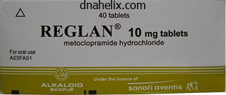
Reglan dosages: 10 mg
Reglan packs: 60 pills, 90 pills, 120 pills, 180 pills, 270 pills, 360 pills

Buy reglan 10mg amexOnly the proximal third of the tibial shaft or only the tibial con dyles are present gastritis leaky gut effective 10 mg reglan. The tibia may be a rectangularly outlined bone with no evident epiphysis; in some circumstances, solely a small bone cap represents the proximal epiphysis. The fibula is positioned usually or rests superiorly and posteriorly in the popliteal house. These deformi ties are usually managed with a Syme amputation and appropriate prosthetic management if the affected person has a functioning extensor mechanism of the knee. If the patient is unable to actively prolong the knee, then treat ment is similar to that for a complete deficiency with a knee disarticulation and subsequent prosthesis. Longitudinal deformities of the ulnar ray (see Plate 447) are sporadic and nonheredi tary and are among the rarest congenital anomalies of the higher limb. Ulnar ray defects are frequently associ ated with malformations of the radial ray (most common) or of the central rays as well. Associated deformities within the shoulder girdle, proximal humerus, or each, can also be present. They include radiohumeral dislocation or synostosis, hypoplasia, partial or complete absence of the ulna, curvature of the radius, ulnar deviation of the hand, fusion of carpal bones, congenital amputation at the wrist, and oligo dactyly with or without syndactyly. Func tional testing of limb position, power, and stability helps to decide the most effective therapy. In common, surgi cal treatment is reserved for the hand anomalies associ ated with ulnar deficiencies. Function may be improved with surgical launch of syndactyly, internet deepening, and thumb reconstruction or pollicization. Occasionally, in partial ulnar defects with significant instability of the elbow, the ulnar remnant could be fused to the radius to provide stability. Humeral part of nonfunctional right prosthesis contains rechargeable battery pack. Total fibular deficiency is probably considered one of the commonest lengthy bone deficiencies and is bilat eral in about 25% of patients. There are usually only three or four toes, and the distal tibial epiphysis is absent or minimal. Treatment consists of an ankle disarticulation amputation and use of an end bearing ankle prosthesis. The tibia is just minimally shortened and the fibula is both quick ened or its distal portion appears regular. Treatment is with a shoe lift, but surgical epiphyseal stapling to arrest progress may be essential. Central ray deficiencies are further classified into typical and atypical subgroups. Typical malformations range in severity from a partial or complete deficit of a phalanx, metacarpal, or carpal bone of the central rays to a monodigital hand. In the syndacty lous kind, which may be partial or complete, the ele ments of the third ray are fused to either the second or fourth digital ray, resembling an osseous syndactyly. The hand has a central cleft of soft tissue and the looks of a lobster claw (see Plate 448). In the polydactylous deficiency, supernumerary bony ele ments are current in the hand, creating a cleft of sentimental tissue and the appearance of a lobster claw. In figuring out therapy for the cleft hand, current function should be thought of. The two opposing digital units are often secure, cell, and quite practical, though not cosmetically enticing. Closure of the cleft consists of reconstruction of the deep transverse metacarpal liga ment. The perform of a monodigital hand could be improved with rotational oste otomy, opponensplasty, use of a simple opposition submit, or a mixture of all three. The most profound longitudinal arrest is phocomelia (see Plate 449), a failure of proximodistal growth. Phoco melia may be complete (the hand or foot is hooked up on to the trunk) or partial (the hand or foot is connected to a deficient, severely shortened limb). The patient with bilateral upper limb phocomelia is unable to place the palms for feeding and toilet activities. Frequently, the issue is further com pounded by associated deformities of the decrease limbs that stop good foot prehension. The joints in phocomelia are usually unstable and hyperextensible due to ligament laxity, and muscle power is decreased. Many sufferers can use the affected limb to control the terminal device or elbow lock in a nonstandard prosthesis, which must be kept so simple as possible to be accepted by the affected person. In partial phocomelia, remedy will not be needed, or one of the following alternatives may be indicated: clavicular transfer to exchange the lacking humerus, use of a nonstandard shoulder disar ticulation prosthesis, hand reconstruction to improve grip or pinch, or remedy to enhance function with the present buildings. In proximal lower limb phocomelia, the ligaments are extraordinarily lax and the tibia slides up and down within the pelvis. In distal decrease limb phocomelia, the foot articulates with the distal femur and is often monodigital. Failure of Differentiation of Parts Failure of differentiation (separation) of parts refers to all deficits in which the essential anatomic models are current however improvement is incomplete. The homogeneous anlage, or primordial, differentiates into the skeletal, dermomyofascial, and neurovascular elements found in a standard limb, but differentiation, or separation, is incomplete. Therefore, this category contains soft tissue involvement, skeletal involvement, and congeni tal tumors. Congenital elevation of the scapula (see Plate 428) and absence of the pectoral muscle tissue are the 2 kinds of failure of differentiation within the shoul der. Soft tissue involvement could also be manifested by aberrations of the long flexor, extensor, or intrinsic muscle tissue within the higher limb. Failure of skeletal differentiation can lead to either dislocation or synostosis of the humeroradial, humeroulnar, proxi mal, or distal radioulnar joint. Synostosis of the proxi mal radioulnar joint, probably the most extreme elbow deformity in this class, is genetically decided and infrequently related to synostosis elsewhere in the body. Surgi cal correction could also be indicated if flexion/extension or pronation/supination deformities that interfere with perform are current. Failure of differentiation can happen in both the skeletal or gentle tissue parts of the carpus, metacarpals, or fingers. In symphalangism, an intermediary joint within the digit is missing, most commonly the proximal interphalan geal joint. Symphalangism of the distal interphalangeal joint is rare and nearly never seen within the thumb. If ankylosis is established, the deformity can be handled with implant arthroplasty or with osteotomy and fusion of the joint in a practical position. Syndactyly, one of the two most common malforma tions within the hand, is commonly bilateral and can involve two or more digits, normally the middle and ring fingers. In some sufferers, solely the gentle tissues are fused (simple syndactyly); in different sufferers, the nails and bones are joined as well (complex syndactyly).
Syndromes - Phlebitis (vein inflammation)
- Intussusception (part of the intestine folds in on itself)
- Antifungal medicines such as fluconazole (taken by mouth) or amphotericin (given by injection) can treat candida infection.
- Mouth pain - severe
- Screen for diseases, such as high blood pressure or diabetes
- 14 to 18 years: 11 mg/day
- Children 5 - 6 years old: 75 - 115 beats per minute
- Diarrhea
- The time it takes you to begin urinating
- Nasopharyngeal culture

Generic reglan 10 mg on-lineCongenital hyperextension and/or dislocation of the knee gastritis symptoms how long does it last reglan 10 mg on line, though uncommon, is an orthopaedic emer gency when it happens. At start, the knee may be merely hyperextended (genu recurvatum) or, within the extreme form, utterly dislocated, with the tibia displaced anterior and lateral to the femur. Dislocations are typi cally bilateral and associated with "syndromic" patterns, corresponding to Larsen or EhlersDanlos syndromes. In an otherwise normal youngster, the dislocation is believed to outcome from an intrauterine place (frank breech presentation), by which the ft of the fetus are locked beneath the person dible or within the axillae. The knee seems "back ward" and hyperextended, with the examiner typically capable of further extend the leg until it nearly touches the chest. The medial hamstring muscle tissue are sometimes dis positioned ahead, anterior to the axis of the knee, thus functioning as knee extensors. The patella may be dis positioned laterally, and the femoral condyles are promi nent posteriorly. Radiographs reveal extreme genu recurvatum with malalignment of the tibia and femur, with a spectrum of findings starting from genu recurvatum to full anterior dislocation. Defor mity of the epiphyses of the distal femur and proximal tibia may be seen in untreated older kids. Within a couple of hours after delivery, the limb must be passively stretched to bring the knee steadily into a flexed place. In most sufferers, the knee can be manipulated into slight flexion (30 degrees) and splinted in this position. The splint should be changed often, with stretching and passive vary ofmotion workout routines continued till the knee may be flexed to approximately 90 levels. A removable splint may be then be used for an additional 2 to 3 months to keep position. The numerous causes of the inequality include the following: � Congenital and developmental anomalies with terminal limb deficiencies (see Plate 332): hemi hypertrophy or hemiatrophy, KlippelTr�naunay Weber syndrome, Maffucci syndrome, posterior bowing of the tibia, proximal femoral focal defi ciency, congenital brief femur, enchondromatosis � Paralytic issues: poliomyelitis, encepha lopathy. A frequent technique is to place standing blocks of measured thickness beneath the short leg to level the pelvis. Radiographic techniques, utilizing a metallic ruler on the movie, include a oneexposure approach during which a single exposure is made of each entire lower limbs. The oneexposure approach could produce magnification on the ends of the lower limbs owing to the impact of paral lax. A extra accurate methodology includes three successive exposures of the hips, knees, and ankles on one lengthy movie (see Plate 433). Technique for radiographic measurement by the three-exposure approach (scanogram). The quantity of progress remaining and therefore the appropriate timing of an epiphysiodesis to equalize leg lengths can be calculated with the chart devised by Green and Anderson, by the arithmetic methodology of Menalaus, or by the Moseley straightline graph (see Plate 434). The Green and Anderson growthremaining chart is used to estimate the results of an epiphyseal arrest pro cedure on the distal femur and proximal tibia at varied skeletal ages. The arithmetic technique of Menalaus assumes that boys close their progress plates at a median age of sixteen whereas girls close their progress plates at a mean age of 14. The Moseley straightline graph helps determine the estimated lengths of the long and brief bones at matu rity, the discrepancy at maturity, and when the most effective equalization procedure ought to be performed. Regular followup is necessary to determine if the discrepancy is progressive and whether or not conservative measures. Surgical procedures for leglength discrepancy embody (1) shortening of the long facet by arresting or retarding epiphyseal growth or resecting a section of bone; (2) femoral, tibial, or transiliac lengthening of the brief facet; (3) combined shortening of the long side and lengthening of the short aspect; and (4) prosthetic fitting. The open technique of Phemister involves eradicating an oblong block of bone at the medial and lateral borders of the expansion plate. The introduction of improved clarity of intraoperative radiographic picture intensification has facilitated using a closed method, percutaneous epiphysiodesis. A very small incision is remodeled a Steinmann pin placed medially to laterally within the airplane of the expansion plate. A cannulated reamer is placed over the pin and used to begin elimination of the growth plate, which is accomplished by energy drilling or curettage or both. Viscous lidocaine and a radiographic distinction medium are injected into the defect, and the limb is rotated underneath the picture intensifier to determine the adequacy of the procedure. Morbidity is type of low, and the scar is much more acceptable to sufferers than that of open epiphysiodesis. If development is to be resumed, the staples should be removed before progress of the epiphysis has ceased. After the staples are eliminated, a rebound phenome non, or initial development spurt, may occur, followed by continuation of development on the normal price. A beforehand stapled epiphysis normally closes a couple of months prema turely, which tends to compensate for the spurt in growth. Although there are many technical issues related to the stapling process, the theoretical advantages of stapling-such as the power to control angular and size deformities-make it a worthwhile consideration. Resection of bone from the longer limb may be per formed to appropriate leglength discrepancy in skeletally mature sufferers and may simultaneously right any associated angular or rotational deformities. The danger of extreme shortening is muscle weakness, which could be manifested in the femur as a knee extension lag as a end result of decreased quadriceps strength. Open epiphysiodesis Straight- and right-angled curets of varied sizes used for full removing of peripheries of development plate (anterior view with knee and hip in flexion). The discrep ancy in a skeletally immature baby must be larger than may be corrected with epiphysiodesis of the long limb, which by conference has been considered to be approximately 5 cm. Muscle energy should be suffi cient so that little energy is lost by lengthening. However, even gradual lengthening could trigger several systemic complications, together with transient hyperten sion, anorexia and weight reduction, and emotional lability. The technique of limb lengthening often recognized as distrac tion osteogenesis was introduced by Ilizarov in 1951 (see Plate 436). After subperiosteal division of the bone at the diaphysis or metaphysis (corticotomy) without disturbing the medullary canal, the bone fragments are fixed above and beneath with an external fixation system. The Ilizarov gadget incorporates metallic rings that encir cle the limb and connect to the bone with thin metal wires or half pins. The De Bastiani system, known as a dynamic axial fixator, is a inflexible telescop ing bar that attaches to one side of the limb with screws (see Plate 436). Rectangular bone plug incorporating progress plate resected from both sides of distal femur. Growth plate drilled and curetted and hole crammed with cancellous bone from above and below. Growth retardation epiphyseal stapling Bone plug reversed and impacted into its bed. After percutaneous or open corticotomy, Ilizarov system secured with wires or pins passing via bone.
Cheap reglanHomologous to corpora cavernosa of the penis diet during gastritis attack buy 10mg reglan amex, the clitoris is about 2 cm lengthy and has two crura containing erectile tissue that end as a rudimentary glans clitoris. A dense connective tissue capsule with an intervening, incomplete septum covers the crura. Erectile tissue of the clitoris consists of a plexus of thin-walled venous channels that distend throughout sexual stimulation. Loose connective tissue and isolated easy muscle cells are associated with these channels. Many nerve fascicles are within the connective tissue; the mucous membrane that covers the clitoris externally incorporates many sensory nerve endings. The vagina and urethra open into the vestibule, which is lined by stratified squamous epithelium. Near the clitoris and urethra, several minor vestibular glands (which resemble male glands of Littr�) secrete mucus. Two bigger tubuloalveolar glands, the major vestibular glands (of Bartholin), open on the internal surface of the labia minora. Connective tissue septa Full-term placenta Amnion enlarging and encircling endodermal tube and fetal mesoderm. B H Amnion completely encircling early fetus which is connected only by the physique stalk. Its spongy architecture is as a end result of of many intently packed chorionic villi and blood-filled intervillous areas. The disc-shaped organ is 1525 cm in diameter and 2-3 cm thick; it weighs 400-600 g at term. The placenta serves many important capabilities associated to physiologic exchanges between mom and developing embryo or fetus, such as trade of gases, electrolytes, and metabolites between maternal and fetal blood. Maternal antibodies are transmitted to the fetus, and the placenta produces several hormones including estrogens, progesterone, and human chorionic gonadotropin. The maternal component of the placenta is the decidua basalis of the endometrium, which is the modified stratum basale in which the embryo is implanted. The fetal part, fashioned from the chorionic sac surrounding the embryo, consists of the chorionic plate and its branching chorionic villi that reach from the chorion like branches of a tree. The tips of the villi are connected to the decidua and, by 6 weeks, branches are formed with free tips, which create a villous spongework. Many chorionic villi finish freely; others fuse with the decidua as anchoring elements. The villi provide a large surface area in touch with maternal blood for nutrient change. At eight weeks (A), placental villi are narrow and coated by two distinct cell layers. By 12 weeks (B), the double trophoblast cell layer is less apparent, and reduced numbers of cytotrophoblasts appear singly, not as a steady layer. Thin-walled fetal capillaries (Cap) are outstanding, most lying in the middle of each villus. By 20 weeks (C) and at time period (D), the many fetal capillaries with enlarged lumina are extra peripherally situated, mendacity near the syncytiotrophoblast layer. Each villus is fashioned from two epithelial cell layers derived from the trophoblast of the embryo, which are carefully related to extraembryonic connective tissue. An internal single layer of cytotrophoblasts, or Langhans cells, consists of cuboidal epithelial cells with light-staining cytoplasm and distinct cell boundaries. They give rise to a steady superficial layer of bigger syncytiotrophoblasts, which stain darker and have ill-defined cell boundaries. Trophoblast cell layers initially type proliferating villous cords that invade the endometrium and destroy the partitions of coiled arterioles and venules of the endometrial stroma. Extravasated maternal blood creates irregular intervillous areas in eroded decidual tissue, circulates in these spaces, and bathes the chorionic villi. The core of each villus consists of loose mesenchymal connective tissue containing fetal capillaries, fibroblasts, and isolated smooth muscle cells. Macrophages, generally identified as Hofbauer cells, are also current in villi and turn out to be extra quite a few throughout gestation. In the second half of pregnancy, cytotrophoblasts progressively disappear, and a skinny layer of multinucleated syncytiotrophoblasts stays on villi surfaces. In the third trimester, local bulges of syncytiotrophoblast nuclei, referred to as syncytial knots, are common. Fetal capillaries in the stroma of each villus obtain blood from umbilical arteries and drain into venules that ship blood to umbilical veins. In placenta accreta-a potentially life-threatening condition to the mother-partial or full absence of the decidua basalis ends in placental adherence on to the myometrium. A fetal capillary (Cap) is intently related to a pale-stained cytotrophoblast (Cy). Microvilli project from the syncytiotrophoblast floor into the intervillous house (to the left). It is lined by an attenuated, continuous endothelium (En) with tight junctions (circles). It consists of the continuous endothelium of the fetal capillary and its basal lamina, a layer of cytotrophoblasts and its adjacent basal lamina, and a layer of syncytiotrophoblasts uncovered to maternal blood. All substances that cross this barrier for gasoline trade, waste elimination, and transport of electrolytes, glucose, and different substances traverse syncytiotrophoblasts. Syncytiotrophoblasts carry out many capabilities, corresponding to endeavor passive and facilitative diffusion, energetic transport, receptor-mediated endocytosis of immunoglobulins, and exocytosis for secretion. These multinucleated cells have ultrastructural features in widespread with secretory, metabolically lively, and absorptive epithelia. Syncytiotrophoblasts arise from cytotrophoblasts, which have a standard complement of organelles. Their plasma membranes interdigitate with these of neighboring cytotrophoblasts and possess desmosomes at lateral margins of adjoining cells and at surfaces in contact with syncytiotrophoblasts. The adventitia, which is a traditional characteristic of most arteries, is replaced by mucous connective tissue of Wharton jelly. In the relaxed state, the lumen of the vein is usually larger than that of the arteries. The prominent tunica media incorporates an interlacing community of round easy muscle cells. It incorporates two umbilical arteries and one umbilical vein coiled around each other in Wharton jelly, a matrix of embryonic connective tissue that has a mucous consistency. This extracellular matrix has an interlacing community of delicate collagen fibers and is wealthy in hyaluronic acid and chondroitin sulfate. It contains stellate or fusiform cells that resemble mesenchymal cells but lacks different blood vessels, lymphatics, and nerve fibers. Via particular staining methods, small autonomic nerves may be seen in the proximal end of the wire. A single layer of cuboidal epithelium, which is derived from the liner of the amniotic cavity and is called amniotic epithelium, covers the twine.

Safe 10 mg reglanThe inner substance of the cornea additionally arises from mesenchyme gastritis diet chocolate discount 10mg reglan with amex, however the anterior floor is epithelium derived from ectoderm. It is brought on by a mutation within the long arm of chromosome 13 (13q14), which finally ends up in an abnormal or absent tumor suppressor gene. Surgical removing of the tumor and enucleation (removal of the eye) are frequent treatments, but new chemotherapy brokers that may cross the blood-ocular barrier, mixed with laser and cryotherapy, provide favorable results. The corneal stroma contains collagen fibers interspersed with modified fibroblasts derived from neural crest ectoderm and generally recognized as keratocytes; clear areas are fixation artifacts. The corneal endothelium-simple cuboidal epithelium-rests on Descemet membrane (arrows). It occupies one fifth of the ocular surface, with its radius of curvature lower than that of the relaxation of the eyeball. Basal cells are polygonal, but the most superficial cells, which retain nuclei, are flattened. The epithelium continuously replicates, and it regenerates in response to wear and tear. Its wealthy sensory nerve supply (from the ophthalmic department of cranial nerve V) is sensitive to touch and ache. Deep to the epithelium is Bowman membrane-a distinguished basement membrane, 8-15 mm thick- that binds epithelium to underlying connective tissue. A distinctive pattern of collagen fibers-regularly arranged, parallel in every layer and at right angles in successive layers-contributes to transparency of the cornea. Simple cuboidal epithelium-misnamed corneal endothelium-lines the posterior surface. Its free (apical) floor is directly exposed to aqueous humor in the anterior chamber. Being avascular, the cornea is immunologically privileged and a good candidate for transplants. The boundary between cornea and sclera (white of the eye) is an abrupt transitional zone, the limbus, where mucous membranes overlaying the sclera (bulbar conjunctiva) and underside of the eyelid (palpebral conjunctiva) be part of the anterior corneal epithelium. Lamellae of often arranged collagen imparts eosinophilia to the corneal stroma. Flattened nuclei of keratocytes (arrows) are aligned in rows between the lamellae. Collagen fibrils within one lamella are parallel to one another, whereas fibrils in the adjacent lamella are oriented at right angles. The distinctive axial periodicity of kind I collagen is shown when the fibrils are sectioned within the longitudinal plane (Upper Right). Fibrils in every lamella are organized at roughly right angles relative to fibrils in adjacent lamellae. Modified fibroblasts derived from the neural crest, referred to as keratocytes, are stellate-shaped cells with numerous dendritic processes that form a syncytium by way of gap junctions. Their cytoplasm additionally houses corneal crystallins that assist reduce backscatter of sunshine, also contributing to corneal transparency. A precision surgical instrument, often recognized as a microkeratome, first creates a thin (80-200 mm) round flap of corneal tissue consisting of outer epithelial and stromal layers. A computer-controlled excimer laser then reshapes the cornea by vaporizing small amounts of stroma in a finely controlled method. The dilator pupillae muscle forms the inside layer of the heavily pigmented epithelium that covers the posterior floor. The stroma-loose connective tissue-contains many blood vessels, flattened fibroblasts, and melanocytes. A double layer of heavily pigmented epithelium is in contact with the posterior chamber. Its free end is suspended in aqueous humor between the cornea and lens; its root is steady with the ciliary body. Its central adjustable aperture is the pupil, whose opening and thus the amount of sunshine reaching the retina, it regulates. Its anterior floor, which contacts the anterior chamber, has, instead of epithelium, a discontinuous layer of stromal cells: a mix of fibroblasts and pigment-containing melanocytes. Spaces between the cells allow fluid from aqueous humor to percolate into the stroma. The stroma is richly vascularized, and most vessels have a corkscrew shape to adjust for size changes in the iris. The number of melanocytes in the stroma and the quantity of melanin in their cytoplasm principally determine eye shade. A double layer of pigmented cuboidal epithelium, continuous with that of the ciliary body, covers the posterior surface. The superficial layer of those cells is in touch with aqueous humor within the posterior chamber. The inner layer is manufactured from myoepithelial cells, which form the dilator pupillae muscle. Postganglionic nerve fibers of the sympathetic nervous system stimulate the cells to contract, which causes pupil dilation. In the stroma, near the pupillary margin, lies the involuntary constrictor pupillae muscle, a flat ring of circumferential smooth muscle, about 0. Cornea Anterior chamber Corneoscleral limbus Canal of Schlemm Trabecular meshwork Lens Ciliary processes Pupil Iris Zonular fibers Ciliary muscle Conjunctiva Sclera Choroid Retina Ora serrata EyeandAdnexa 437 Anatomy of the lens. External look Embryonal nucleus Fetal nucleus Infantile nucleus Adult nucleus Cortex Epithelium Capsule Anterior phase of the attention exhibiting the topography of the lens. Refractive media of the eye-cornea, lens, aqueous and vitreous humors-help focus images on the retina. The rest of the lens is manufactured from concentric, transparent refractile fibers, most missing nuclei (arrows). One row of simple cuboidal epithelial cells with spherical nuclei rests on an exterior capsule. It is held in a reasonably fixed place by zonular fibers (from the ciliary body) and the vitreous body behind it. Lens fibers, the principle cells of the lens, are elongated columnar epithelial cells with distinctive cytoplasmic proteins (crystallins), filensin interme- diate filaments, and a degenerated nucleus. Enveloping the lens is a homogeneous capsule that corresponds to a thick basement membrane containing a network of collagen fibrils. Underneath it, the anterior half of the lens has a easy cuboidal (lens) epithelium, the germinal zone. The lens epithelial cells within the equator undergo mitosis and differentiation all through life. Anterior chamber Corneal part Slit lamp beam Anterior capsule Adult nucleus Embryonal nucleus the creating lens earlier than start displaying orientation of lens fibers. These processes, like a ball and socket (circle), hold upper and lower surfaces of fibers together. Scanning electron microscopy of the lens after freeze fracturing reveals that fibers are hexagonal prisms about 10 mm lengthy, 10 mm wide, and a couple of mm thick. Most fibers are in rows, organized concentrically and parallel to the lens surface.

Best order reglanAround nuclei is commonly a halo sample gastritis symptoms pain order reglan american express, which is due largely to glycogen that by light microscopy often appears washed out. Short basal infoldings of plasma membrane (arrows) enhance floor space and comprise ion pumps. Tight junctions (circle) link apicolateral cell borders and seal the lumen from the extracellular space. Dark cells, which may play a more lively role in urine acidification, have extra organelles, apical vesicles, and basal infoldings than do light cells. Apical surfaces of darkish cells even have more numerous and stubby microvilli; light cells may bear a single cilium. Tight junctions connect both cell types; basal plasma membranes rest on a thin basement membrane. Thus, not solely are collecting ducts conduits, but they also play a task in concentrating urine and regulating acid-base steadiness. Somite Intermediate mesoderm (nephrotome) Pronephric tubule Pronephric duct Ectoderm Pronephric duct forming UrinarySystem 375 Dorsal aorta Glomerulus Coelom Gut Pronephric tubules degenerating Mesonephric duct Mesonephric tubules in nephrogenic tissue Ureteric bud (metanephric duct) Metanephrogenic tissue Topography of the pronephros, mesonephros, and metanephric primordium. Somite Dorsal aorta Glomerulus Posterior cardinal vein Mesonephric duct Mesonephric tubule Genital ridge Coelom Gut Cloaca Horseshoe kidney. This common renal fusion defect occurs when intermediate mesoderm that offers rise to the metanephric blastema fails to separate. Both methods arise from mesoderm: At 4 weeks of gestation, intermediate mesoderm separates from successive somites to type segmentally organized nephrotomes, that are just lateral to the genital ridge. They give rise, in a cranial to caudal path, to three successive kidneys-pronephros, mesonephros, and metanephros. The pronephros forms seven pairs of pronephric tubules and a pronephric duct, which extends to the caudal a half of the embryo to attain the cloaca. The vestigial and nonfunctional human pronephros is quickly replaced caudally by the mesonephros, which serves briefly as an excretory organ in the fetus. The mesonephros consists of tubules that fuse with an extension of the pronephric duct, referred to as the mesonephric (wolffian) duct. Successive formation of tubules within the caudal a half of intermediate mesoderm continues for a quantity of weeks, with 16. Primitive renal glomeruli type in the mesonephros between blind ends of tubules and capillaries derived from branches of the dorsal aorta. After mesonephros regression, the metanephros (permanent kidney) appears within the fifth week of gestation. Other than symptomatic remedy, surgical intervention may be undertaken in some circumstances to enhance urine circulate. Mesonephron Mesonephric duct Hindgut Cloacal membrane Cloaca Metanephrogenic tissue Metanephric duct (ureteric bud) Metanephrogenic tissue Capsule Pelvis Major calyx Minor calyx Collecting ducts Nephroblastoma (Wilms tumor). The metanephric duct (ureteric bud) has grown out from the mesonephric duct, near termination of latter in cloaca, and has invaded metanephrogenic mesoderm. Distal ends of collecting ducts join with the tubule system of the nephron creating from metanephric mesoderm. The tubule lengthens, coils, and begins to dip down towards the renal pelvis, as Henle loop; one part of the tubule stays close to the glomerular mouth, as the longer term macula densa. Within metanephrogenic tissue, the bud expands to type a pelvis, which branches into calyces, which, in flip, bud into successive collecting ducts. Proximal convoluted tubule Distal convoluted tubule Macula densa Renal corpuscle Kidney Collecting tubule Henle loop Tumor Ureter Wilms tumor with pseudocapsule and characteristic variegated structure. The loop elongates; renal corpuscle, proximal tubule, Henle loop, distal tubule, and macula densa of mature nephron thus derive from metanephrogenic mesoderm and collecting tubules from the metanephric duct. The bud-an outgrowth of the mesonephric duct-gives rise to ureters, renal pelvis, renal calyces, amassing ducts, and accumulating tubules. These tubules endure dichotomous branching, and by the 20th developmental week, about 10-12 generations of ducts have fashioned. Metanephrogenic tissue from the caudal a part of intermediate mesoderm gives rise to remaining elements of nephrons: proximal and distal tubules, Henle loop, and Bowman capsule of the renal corpuscle. Terminal branches of collecting tubules are first covered at distal ends by mobile aggregates of metanephrogenic tissue. These aggregates form hollow vesicles that turn into primitive tubules with a central lumen, which then become nephrons. The tubules, lined by easy epithelium, turn out to be covered externally by continuous basement membrane, elongate, and eventually reach their convoluted grownup kind. Epithelium covering distal (free) ends of the tubules turns into flattened and is invaded by a tuft of glomerular capillaries to type a renal corpuscle. The primitive nephron lines up with the amassing tubule and the two fuse to type a passage for urine. They encompass immature and mature mesenchymal tissues mingled with abortive glomeruli and renal tubules. The irregular stellate contracted lumen is lined by urothelium, which rests on a lamina propria of unfastened connective tissue. Two layers of loosely organized clean muscle are easily seen in the higher part of the ureter; three layers happen in its decrease half. In this form of urinary incontience, lack of small amounts of urine is commonly related to coughing, sneezing, or straining. Epithelium in the upper a half of the ureter consists of two or three cell layers; it progressively modifications to four or five layers within the lower third. Epithelium thickness is dependent upon the diploma of distention and varies markedly from thinner within the distended state to relatively thick in the collapsed (or empty) state. Plasma membranes of essentially the most superficial epithelial cells, which are in direct contact with the lumen, have an accordion-like pleating capability. Also, the epithelium is nearly impermeable to movement of water or ions, so the concentration of urine remains fairly constant as it passes down ureters into the bladder. Second, an underlying cellular and fibrous lamina propria helps the epithelium. Epithelium and lamina propria together represent the mucosa (or mucous membrane) of each ureters and bladder. Third, a muscularis externa consists of easy muscle organized in layers that are opposite in orientation to these within the digestive tract wall. The fourth, outer layer is an adventitia (or serosa), which consists principally of loose connective tissue with autonomic nerves and plexuses, blood vessels, and lymphatics. Normal neurologic management of bladder and urethral perform entails smooth muscle receptors for neurotransmitters of both sympathetic and parasympathetic nervous methods. Stress incontinence is a standard postsurgical complication of radical prostatectomy in men. Interlacing smooth muscle bundles occupy the muscularis externa, which is invested by an outer adventitia. Calculus impacted in upper finish of ureter with resultant hydronephrosis; additional stones in kidney and renal pelvis. Lumen Urothelium Dilated ureter Peritoneum reflected Stone in decrease end of ureter in process of removal through ureterostomy. The wall is manufactured from mucosa, which consists of urothelium that rests on a lamina propria of pretty dense collagenous and elastic connective tissue.
Clarified Honey (Honey). Reglan. - What is Honey?
- Wound healing. Applying honey preparations directly to wounds or using dressings containing honey seems to improve healing.
- Cough. Taking a small amount of honey at bedtime appears to reduce the number of coughing spells.
- Dosing considerations for Honey.
- Burns. Applying honey to the skin seems to help improve healing.
- How does Honey work?
- Sunburn, foot ulcers caused by diabetes, asthma, allergies, breaking up thick mucus secretions, diarrhea, digestive tract ulcers, and cataracts.
- What other names is Honey known by?
Source: http://www.rxlist.com/script/main/art.asp?articlekey=96721

Buy cheap reglan lineBiopsy findings of colloid-containing follicles typical of thyroid carcinoma (H & E stain) gastritis symptoms buy reglan 10mg line. Biopsy results indicated adenocarcinoma of kidney, which was confirmed by renal arteriogram and intravenous pyelogram (section below stained with H & E). Radiograph revealed destructive lesion suggestive of osteosarcoma, but histologic findings indicated adenocarcinoma. Elevated acid phosphatase stage and outcomes of digital rectal examination confirmed origin (H & E stain). Colon cancer is the subsequent most common primary malignancy to metas tasize to bone after the aforementioned 5. The most typical sites of involvement for bone metastases are these containing hematopoietic marrow. The uncommon metastasis to cortical bone or to the bones of the palms or ft suggests seeding from a major lung carcinoma. In youngsters, metastatic skeletal tumors are often secondary to neuroblastoma in those younger than 2 years old or to lymphoma and Ewing sarcoma in older youngsters. Metastasis after age forty is usually sec ondary to the primary carcinomas talked about earlier. Purely osteolytic metastases are generally from lung, kidney, thyroid, or colon most cancers. Metastatic bone tumors from kidney or thyroid carcinoma are normally hypervascular, and preoperative embolization must be considered. In sufferers with a history of carcinoma, the presence of a solitary tumor necessitates a seek for different sites of skeletal involvement. Even with no identified primary tumor, an isolated lesion in someone older than 40 is normally a metastasis and less probably myeloma or lymphoma. Primary sarcomas in patients older than forty are much much less doubtless than metastatic disease, myeloma, or lym phoma. For a number of metastatic lesions, chemotherapy, hormonal manipulation, and palliative radiation remedy are therapeutic options. Surgery is carried out for most pathologic fractures and for some pathologic lesions which are believed to have excessive chance of fracture (impending fractures), such as these with over half of the diameter of the bone destroyed or with bone corti cal destruction over three cm in length. Prognosis for patients with skeletal metastases is poor, although sufferers with isolated bone metastases particularly with breast most cancers and renal cell most cancers can reside for a number of years. It could occur at any age, first showing as an asymptomatic but slowly enlarging soft tissue mass, often adjoining to fibrous or fascial structures and fre quently in extracompartmental soft tissues. The palmar and plantar elements of the arms and ft are frequent sites of involvement. Treatment of desmoids has historically been by excision or resection, but primarily based on the extraordinarily excessive recurrence rate different less invasive strategies corresponding to tamoxifen and nonsteroidal anti inflammatory medicine have met with some early success. Usually nonoperative remedy ought to no much less than be attempted with surgical procedure reserved as a last resort. Fibromatosis of buttock with extensions into gluteus maximus muscle Hemangioma the term fibromatosis refers to a number of fibrous tumors that, although benign, are significantly more aggressive than solitary fibroma. These lesions typically develop in the proximal limbs or the trunk and are also called stomach and extraabdominal desmoid tumors, depending on whether or not or not they involve the abdomi nal wall. Although usually superficial, the tumors are locally invasive, regularly involving adjacent neurovas cular constructions. Although the lesions normally develop adjacent to or even adherent to bone, bone scans are often cold. Histologic examination reveals scattered spindle cells enmeshed in heavy strands of mature collagen. Because the extent of involve ment and the aggressiveness of fibromatosis are often underestimated, the surgical margins achieved may be insufficient as with desmoids tumors. Recurrences are troublesome to distinguish from the scar ring of earlier excisions, thus making subsequent exci sion much more troublesome. Adjuvant radiation remedy may scale back the recurrence fee after marginal, and even after intracapsular, excision however has its personal threat and should even cause malignant degeneration of these benign lesions. Sometimes, hemangioma is congenital, appear ing as a solitary tumor that infiltrates local tissue and, like fibromatosis, might involve adjacent neurovascular constructions. The commonest form is a tumor with infiltrative margins composed of both massive and small vessels. On physical examination, elevat ing the affected physique part above the extent of the guts might noticeably scale back the dimensions of the mass by emptying venous blood within the lesions. However, hemangiomas regularly recur after surgical resection; due to this fact, injection by interventional radiology or vascular surgery has been successful and may typically be attempted, with resection as a last resort. It happens in adults and is usually gentle, slowly enlarg ing, and asymptomatic and normally located superficially within the subcutaneous tissues. Lipomas that come up in deep tissues could turn into remarkably large and have an increased chance of being an atypical lipoma. Radiographs may also show cal cification in areas of necrosis and metaplastic bone or cartilage inside lipomas. Neurofibromas also occur at the side of scoliosis, congenital pseudarthrosis of the tibia, and gigantism of a limb. These lesions originate from a nerve, and due to this fact resection will normally require no less than slicing nerve bundles. In contrast, schwannomas are benign tumors of the nerve sheath and might fre quently be excised without demonstrable damage to the adjacent nerve. If a neurofibroma is excised, the nerve, or a minimum of the nerve fascicle of origin, might need to be excised. Section reveals unfastened, fibrillar, wavy neural strands attribute of neurofibroma (H & E stain). Myositis ossificans is a nonneoplastic reparative or reactive ossification of sentimental tissue that usually occurs after blunt trauma, notably in conjunction with head trauma. It can happen in adolescents as a painless, enlarging mass in the higher arm, thigh, or buttocks. This clinical presentation could erroneously suggest an extraosseous osteosarcoma or gentle tissue sarcoma. Radiographs reveal a round mass with a definite margin of mature ossification and a radiolucent center of immature osteoid and primitive mesenchymal tissue (the reverse of that seen in a malignant tumor). Malignant fibrous histiocytoma is seen in adults and primarily metastasizes to the lungs. Fibrosarcoma of soft tissue the diagnosis of fibrosarcoma is rare (see Plate 625). Lowgrade stage I fibrosarcoma is usually troublesome to distinguish from its benign but aggressive counterpart of fibromatosis (see Plate 623). Histologic characteristics are marked cellular atypia and abundant collagen in a herringbone sample. A lowgrade fibrosarcoma may be confused with aggressive juvenile fibromatosis, a diagnostic issue that affects therapy choices. Large, infiltrating tumor encompassing knee typifies high-grade, delicate tissue fibrosarcoma which will happen at many websites.
Order reglan 10mg onlineA few isolated ependymal cell nests (arrows) are also seen nearby gastritis patient handout reglan 10mg mastercard, and their presence is normal in the adult. The central canal of the spinal wire is normally patent within the child and younger adult however with advancing age often becomes obliterated. The ventricular lumen (*) has a ciliated ependymal lining composed of carefully apposed cuboidal cells, a few of which bear apical cilia (arrows). These cells bear apical microvilli to increase surface space, and most even have motile cilia that project into the ventricular lumen. Characteristic of the ependyma is the presence of apical intercellular junctions between lateral borders of contiguous cells; different kinds of junctions are adherens, tight, and hole junctions. Intracranial lesions, arising from the roof of the fourth ventricle, normally occur in children, whereas spinal wire tumors typically happen in adults. Treatment is determined by neurosurgical intervention to facilitate definitive diagnosis. Postoperative adjuvant remedy consists of radiation of the brain or spinal wire, chemotherapy, or radiosurgery. A causal relationship between these mutations and tumor development has not yet been proved, nonetheless. Hemisection of brain, brainstem, and spinal cord, with the level of part by way of the anterior diencephalon shown. A leaf-like course of covered by easy cuboidal epithelium (Ep) is raised into small floor protrusions known as villi (Vi). The choroid plexus consists of highly branched leaf-like folds of vascularized pia mater coated by a modified ependyma, which is a secretory and ion-transporting epithelium. This simple cuboidal or low easy columnar epithelium rests on a thin basement membrane. A core of unfastened connective tissue of the pia mater accommodates a tortuous community of large fenestrated capillaries which are highly permeable. This course of involves energetic transport of sodium ions and passive diffusion of water. Tight junctions hyperlink lateral borders of epithelial cells, and basal membranes of the cells have many infoldings much like these seen in other ion-transporting epithelial cells. With age, frivolously eosinophilic and calcified concretions, often known as corpora arenacea, may accumulate within the choroid plexus. To acquire fluid, a spinal needle is inserted into the subarachnoid house between lumbar vertebrae L3/L4 or L4/L5. Routine evaluation consists of microscopic examination with Gram stains; exams for glucose; lactate; protein; red blood cell count; and white blood cell rely with differential; and cultures for micro organism, fungi, or viruses. Deeper, ill-defined layers contain various varieties of neurons that make up the remaining cortical grey matter. Small blood vessels (arrows) from the pia mater penetrate the cerebral cortex substance. The surrounding neuropil is an interwoven meshwork of neuronal and glial cell processes. The outer surface is highly folded to enhance the floor space, estimated at about 2000 cm2. Different types of neurons and fibers are organized in horizontal layers, so the cortex seems laminated. Despite regional variations, the cortex sometimes consists of six illdefined layers, which differ in neuronal population density. As many as 5 forms of cortical neurons exist, but pyramidal cells and stellate cells are most numerous. Nerve fibers are oriented tangentially and radially, establish complex intracortical circuits, and transmit impulses at a quantity of synaptic sites. Many neurons make connections with other cortical neurons or project to different areas of the mind and spinal cord. Pyramidal cell bodies, formed like isosceles triangles, vary from 10 to 50 mm in diameter. A massive dendrite initiatives apically, is oriented at right angles to the surface, and branches repeatedly as it climbs to the floor. Emerging from the base of each cell is a single axon that penetrates to deeper cortical layers and enters the medullary white matter. In sure cortical areas, big pyramidal neurons, called Betz cells, have diameters as much as one hundred mm. NervousTissue 121 Purkinje cells (inhibitory) Dendrites of Purkinje cell Molecular layer Purkinje cell layer Granular layer White matter Area of the cerebellum delineated by the rectangle is seen within the micrograph beneath. An antibody to calbindin selectively labels Purkinje cells, in order that their cell bodies, basal axons, and elaborate apical fan-like dendritic tree are clear. It consists of a surface layer of cortex of grey matter and a medullary middle of white matter. Also, the cerebellar cortex most probably incorporates extra neurons than the cerebral cortex. The cerebellar cortex has a remarkably uniform trilaminar organization: an outer molecular layer, an internal layer of granule cells, and a middle monolayer of enormous pear-shaped neurons known as Purkinje cells. Ultrastructural features- soma and apical dendrite-of a Purkinje cell are clearly seen. The perikaryon accommodates ample rough endoplasmic reticulum, free ribosomes, scattered mitochondria, and lysosomes. The surrounding neuropil accommodates neuronal processes of varied dimension interspersed with glial cell processes. They type a single row of uniformly organized, giant neuron bodies on the outer surface of the granule cell layer. Light microscopy reveals a single, vesicular nucleus with prominent Nissl substance in surrounding cytoplasm. By electron microscopy, primary and secondary dendrites are smooth surfaced; small tertiary branches have quick, stubby spines. Each Purkinje cell has greater than one hundred,000 dendritic spines that markedly improve its floor space for synaptic contact. A single myelinated axon tasks from the base of each Purkinje cell and descends to the underlying medullary white region. Granule cells are densely packed, round to oval, small neurons, about 5 mm in diameter. Several brief dendrites project from the base of every granule cell, and one apical axon extends into the molecular layer, loses its myelin sheath, and bifurcates up to 3 mm in each path. Because of their orientation parallel to the floor, unmyelinated axons are often identified as parallel fibers. They establish multiple synaptic contacts with dendritic spines of Purkinje cells. Abnormal activation of a number of sign transduction pathways could lead to tumor formation. Treatment combines surgical resection, craniosacral radiation, and intrathecal chemotherapy.

Discount 10 mg reglan visaNoninvasive strategies to measure bone density have provided a useful definition of osteoporosis: a mass-per-unit volume of usually mineralized bone that falls beneath a populationdefined threshold for spontaneous fracture gastritis symptoms lightheadedness order reglan visa. If web bone resorption exceeds net bone formation, bone mass declines with time (see Plate 2-30). Under these conditions, with a fast rate of bone turnover, the lower of bone mass is speedy. Conversely, with a gradual price of bone turnover, the decline in mass is correspondingly gradual. Similarly, if internet bone formation exceeds net bone resorption (as in normal bone growth and even after longitudinal development ceases), bone mass increases with time. Some potential combos of bone formation/ resorption are proven within the desk within the higher half of Plate 2-30 and graphically offered within the lower half. Example 8 represents regular bone turnover: bone formation and bone resorption are appropriately coupled, resulting in a steady bone mass with no internet change (black bar on center line). This state of bone reworking is seen in the energetic stage of Paget disease (see Section 3, Metabolic Diseases, Plates 3-44 to 3-46). Example 11 exhibits decreased bone turnover fee, but here, too, bone formation and resorption are appropriately coupled, with no internet change in bone mass. Example 1 depicts a state of extreme uncoupling (decreased bone formation and increased bone resorption), which causes a net decline in bone mass over time. Such an imbalance in formation and resorption happens in continual glucocorticoid extra, which may be endogenous (Cushing syndrome) or iatrogenic (see Section 3, Metabolic Diseases, Plate 3-26). Example 2 illustrates elevated bone turnover fee with a normal rate of bone formation however an elevated rate of bone resorption. This state is represented within the decrease half of Plate 2-30 by the black bar beneath the zero line and by the pink dot above the green column, indicating resultant osteoporosis. Mild Normal bone turnover Decreased bone turnover hyperthyroidism, delicate hyperparathyroidism, and a chronic dietary calcium deficiency can cause increased bone turnover, with resorption exceeding formation. In severe hyperthyroidism and hyperparathyroidism, transforming rates can be even greater than those shown in example three, with a correspondingly higher distinction between formation and resorption and infrequently a greater internet lack of bone mass. Stimulation of deposition Weight-bearing activity Growth Fluoride Electricity More (or more active) osteoblasts (B) 2. In a pathologic context, a localized (focal) phenomenon of elevated bone formation and resorption with formation exceeding resorption occurs in the osteoblastic stage of Paget disease. Example 6 illustrates normal bone resorption paired with a rise within the fee of bone formation, which leads to a web improve in bone mass with time. This is typical during normal progress and improvement, especially between late adolescence (when longitudinal development ceases) and roughly age 35 (when peak adult bone mass is reached). Example 7 exhibits the highly uncoupled state of elevated bone formation and decreased bone resorption that end in a internet improve in bone mass. Such a state of transforming is seen within the adult, or autosomal dominant, type of osteopetrosis (see Section three, Metabolic Diseases, Plate 3-43). In this disorder, the decreased rate of bone resorption is due to the relative decline in osteoclastic bone resorption; the speed of bone formation could additionally be regular or increased. This form of uncoupling may be induced by pharmacologic doses of fluoride administered to stimulate bone formation and stabilize bone apatite crystal and render it extra proof against breakdown. Example 9 shows a state of regular bone resorption with decreased bone formation, which leads to decreased bone mass and osteoporosis. This can occur after exposure to a poison or toxin that affects the osteoblasts, as with use of certain chemotherapeutic brokers and continual alcohol abuse. In instance 10, each bone formation and resorption are decreased but bone resorption still exceeds bone formation. This is seen when calcium and vitamin D supplementation helps to diminish age-related bone loss. Example thirteen shows a constructive bone stability ensuing from a standard fee of bone formation and a decreased price of bone resorption. This happens in ladies receiving replacement therapy with calcium and estrogen or calcium and calcitonin quickly after menopause. These agents, administered independently or together, act to decrease bone resorption. The coupling course of then causes bone formation rates to adjust downward accordingly (example 12); eventually, bone mass stabilizes at a a lot decrease rate of bone transforming (example 11). In a extra excessive instance, administration of bisphosphonates to sufferers with osteoporosis nearly eliminates bone resorption charges and, as a consequence of coupling, bone formation charges additionally considerably decline over remedy time. Inhibition of withdrawal Weight-bearing exercise Estrogen Testosterone Calcitonin Adequate vitamin D intake Adequate calcium consumption (mg/day) Child: 400�700 Adolescent: 1,000�1,500 Adult: 750�1,000 Pregnancy: 1,500 Lactation: 2,000 Postmenopause: 1,500 Level of bone mass remains fixed when price of deposition equals fee of withdrawal (osteoblastic exercise equals osteoclastic activity), whether both charges are high, low, or normal. However, in lots of circumstances, early adjustments shall be checked by concomitant stimulation of the opposing bone cell population-stimulation of osteoclasts (resorption) leads to a coupled stimulation of osteoblasts (formation). However, sufficient calcium could be troublesome to obtain in many reasonable diets and has led to fortifying food products with supplementary calcium. On the opposite hand, phosphate is current in nearly all foods and dietary deficiencies are unusual. Accessory elements selling absorption of calcium from the gut embrace an acid pH, a low serum phosphate focus (to avoid exceeding the important solubility product talked about in the first axiom above), and the absence of chelators such as phytate, oxalate, or extreme free fatty acids. The mechanism of phosphate absorption is less selective than that of calcium absorption but also appears to be a minimum of partly depending on the vitamin D metabolites. Because dietary consumption of phosphate varies extensively and absorption is sort of unrestricted, the primary axiom might counsel that people stand poised on the brink of metastatic calcification and ossification because of a high, uncontrollable intake of phosphate. In truth, the renal excretory mechanisms exert a fine-tuned control over phosphate ion ranges. Acting on the level of the gut lining cell (with vitamin D) to increase absorption of calcium three. Acting at the level of the renal tubule (with vitamin D) to improve tubular reabsorption of filtered calcium 4. Acting at the stage of bone (with vitamin D) to increase the inhabitants of activated osteoclasts, which destroy not only the hydroxyapatite crystals but also segments of natural bone matrix, thus releasing each calcium and phosphate ions 5. This is achieved principally by diminishing the osteoclast inhabitants and activity and, to some extent, by reducing gastrointestinal absorption. However, it must be clearly famous that though the second mechanism may be properly developed in avian species and although administration of exogenous, non� species-specific calcitonin could have a profound effect on the skeleton, the natural mechanism in people seems to be too restricted to protect the physique from hypercalcemia. Hyperphosphatemia, or increased focus of serum phosphate, could result in metastatic calcification, particularly in renal failure, for the rationale that critical solubility product may be exceeded even when calcium ranges are normal. Also, gastrointestinal absorption and tubular reabsorption of calcium, and even bone breakdown, are initially reduced, thus diminishing the focus of calcium. The basic axioms and the interactions of the various hormonal and mineral materials discussed here are necessary in understanding the rules that govern and control the homeostatic mechanisms and the alterations that lead to the rachitic syndrome (see Plates 2-28 and 2-29, and Section three, Metabolic Diseases, Plates 3-13 to 3-23). These axioms are distinct, representing chemical, physiologic, and biologic facts. In truth, at the pH of physique fluids and the concentrations of calcium and phosphate ions present in extracellular fluids, the important solubility product is approached and generally exceeded.

Purchase reglan 10 mg without prescriptionSkeletal muscle fibers are elongated chronic gastritis malabsorption purchase 10mg reglan amex, cylindrical cells, 50-200 mm in diameter, with tapered ends. Their cytoplasm, generally known as sarcoplasm, is full of myofibrils, which are cylindrical bundles of myofilaments along the length of the fiber. Each myofibril has a uniform diameter and consists of equivalent repeating units, known as sarco- 4. Sarcomeres are composed of longitudinally oriented thick and skinny filaments and perpendicular Z bands. In longitudinal section, skeletal muscle fibers show transverse striations as a outcome of adjacent myofibrils are in lateral register with each other across the width of the fiber. The larger density of the A bands is due mainly to the presence of thick (myosin-containing) filaments, whereas the lighter density of the I bands is as a end result of of the prevalence of skinny (actin-containing) filaments. In the center of every A band is a lighter H zone (the central a half of thick filaments not overlapped by skinny filaments), which is bisected by a skinny, darkish M band. The width of the I band and H zone in every sarcomere varies and is dependent upon the extent to which the muscle fiber is contracted or stretched. Eosinophilic staining traits and punctate look are due to the contractile proteins, which constitute much of the sarcoplasm of each cell. Nerve fascicles (Nerve) of varied sizes and capillaries (C) abound in surrounding endomysium. Mitochondria (Mi) occur singly between myofibrils or in clusters underneath the sarcolemma. The peripheral nuclei (N) of muscle fibers are usually euchromatic and infrequently comprise nucleoli. A small nerve fascicle and collagen (Co) are within the perimysium between muscle fibers. In transverse sections stained with hematoxylin and eosin (H&E), the sarcoplasm of every fiber is very eosinophilic and appears punctate due to tightly packed myofibrils. Myofibrils constitute the majority of every fiber and principally comprise the main contractile proteins myosin and actin, which make up the myofilaments. The surrounding endomysium helps a wealthy vascular and nerve provide, which consists of capillaries and nerve fascicles near the muscle fibers. Myofibrils are rounded to irregular in shape, and the intervening, intermyofibrillar sarcoplasm accommodates quite a lot of different organelles, mitochondria being probably the most conspicuous at low magnification. In some instances, electron microscopic analysis of plastic-embedded sections might yield additionally useful diagnostic data. The densities correspond to junctional end-feet and provides a scalloped look to the triad junction. Intermyofibrillar mitochondria (Mi) are often oriented in the longitudinal aircraft with facet branches (*) that course transverse to the level of the I band. Each sarcomere is bounded by Z bands (Z) and is shaped by the exact and orderly alignment of myofilaments. T tubules, a membrane system external to the muscle fiber on the junction of A and I bands, penetrate the fiber inside at regular intervals, principally in a transverse airplane. They are aligned at strategic websites within muscle fibers; they normally occur at I band levels or in aggregates at the periphery of the fibers. Their density, location, and distribution in muscle fibers rely upon muscle fiber type. Interdigitation of thick and thin filaments allows sarcomere contraction, which is greatest explained by the sliding filament mannequin by which actin filaments slide along myosin filaments. During contraction, both units of filaments retain their regular length, A bands remain unchanged in length, I bands shorten, and H zones are narrowed. Schematic displaying interplay of myosin and actin filaments at rest and through contraction. The Z band is drawn closer to the edge of the A band by the sliding of filaments, and the I band area narrows. A third filament system is manufactured from single molecules of titin, one of many largest identified proteins, that connects the Z band to the M band. Titin contains elastic parts that act as molecular springs and contribute to the passive elasticity of muscle. Nebulin, one other large protein, spans the length of skinny filaments and forms a fourth filament system in skeletal muscle. At Z bands, thin filaments, nebulin, and titin are anchored to the protein a-actinin. In half of the thick filament, myosin heads are oriented in a single course; these within the other half are in the other way. Each thin filament, about 1 mm long and 5 nm in diameter, consists of a double helix of filamentous actin. Two proteins related to actin, tropomyosin and the troponin complicated, reply to varying calcium ion concentrations by performing as a swap to enable or disable the interplay and formation of cross bridges between actin and the myosin heads. Myosin heads bind to actin and draw the thin filament a brief distance previous the thick filament. Then, linkages break and re-form farther along the skinny filament to repeat the method. It might arise in any muscle group by sudden exposure to chilly temperature but normally improves after the muscles have been warmed up with brief train. Myotonia congenita (or myotonic muscular dystrophy) is a genetic disorder that usually begins in infancy or early childhood. Subsequent interruption in Cl- move triggers prolonged muscle contractions and stiffness. Clinical analysis is by genetic testing, electrodiagnostic procedures, and muscle biopsy. Although the disease severity differs amongst people, physical therapy and other rehabilitative procedures could facilitate muscle operate. The part passes by way of totally different components of A bands of sarcomeres and shows an orderly arrrangement of myofilaments in every region (A, the thick and skinny filament overlap zone; H; M). The inset reveals the sq. lattice pattern of the Z band (Z) and related thin filaments in close by I band (I). Myofilaments in cross part are electron-dense, punctate profiles; the diameter of thick filaments is more than twice that of skinny filaments. Cross sections of I bands show solely skinny filaments, whereas A bands show both thick and thin filaments, which seem as hexagonal networks with myosin mounted on the M band. Where the 2 sets of filaments overlap, the networks mesh so that each thick filament is within the 4. The interval between thick and thin filaments in the double hexagonal array is 10-20 nm. M bands, which are within the heart of the H zones, show thick filaments with fantastic interconnections. Z bands in cross part show a typical square lattice pattern, and skinny filaments within the quick neighborhood are organized in a daily array.

Discount reglan on lineThe pelvis is brief and broad with relatively extensive gastritis operation order reglan in india, nonflaring iliac wings, small and deep greater sciatic notches, and hori zontal superior margins of the acetabulum (champagne glass shape). Birth weight and size could additionally be low regular, and the short stature may not be acknowledged until the affected person is 2 or three years of age. The typical appearance is a thick, stocky physique with a comparatively long trunk and disproportionately short limbs, making the upper physique phase longer than the decrease physique section. The face is also regular with no midfacial hypoplasia or despair of the nasal bridge. Ligamentous laxity is often gentle, and vary of movement within the elbow, particularly extension and supination, is commonly limited. The trunk generally reveals mildly exaggerated lumbar lordosis with a sacral tilt and a barely protuber ant abdomen. Generalized shortening of the long bones with gentle metaphyseal flaring is most notable at the knees. The height of the vertebral our bodies is regular, and the dorsal borders are only mildly scalloped. The undersulfation of proteoglycans within the collagen matrix impairs the response of cells to fibroblast growth factor and limits endochondral progress. A deadly variant is characterised by a decrease start weight than in the basic type, radiographic proof of overlapping joints, dislocation of the cervical spine, and congenital heart illness. Formerly, sufferers with related however less extreme signs had been thought to have a variant type or a unique condition. The variations, extra obvious than real, have been because of variable phenotypic expression. A distinctive group of malformations is evident at birth, with further traits appearing later. In the new child interval, the pinnacle seems regular, but many sufferers develop a characteristic facial look with a slim root and broad midportion of the nostril, lengthy and broad lip philtrum, and sq. jaw. The prominent space across the mouth, coupled with the opposite charac teristic facial options, gave rise to the now out of date term cherub dwarf. Capillary hemangiomas are frequent within the midforehead but fade or disappear with age. Abnor malities of the palate are seen in 50% of patients and embody full, partial, or submucous clefts, bifid uvula, or double uvula with a median longitudinal ridge. These palatal abnormalities-and probably laryngeal defects-produce the characteristic soft rasping or hoarse voice. In 80% of sufferers, the ears swell within the first few days or perhaps weeks after birth, giving the looks of acute irritation. The swelling subsides spontaneously in 4 to 6 weeks, leading to a cauliflower ear. Reduced top is primarily as a outcome of rhizomelic brief ening of the limbs and is additional augmented by flexion contractures of the joints, especially the hips and knees. Partial and complete joint dislocation can be frequent, particularly within the shoulders, elbows, hips, and patellae. The dysplastic hip adjustments, coxa vara, and hip disloca tion mix to produce a grossly irregular gait. The hypermobile thumb and deformed first metacarpal create an abducted hitchhiker place. Kyphosis of a variable diploma accompanies the scoliosis, and the resultant deformity additional reduces peak. Spinal changes, especially cervical kyphosis, might trigger catastrophic neurologic problems. Devel opment of the epiphyses is delayed and irregular, and stippling has been observed. The epiphyses of the Progression to typical cauliflower deformity proximal femurs, absent at start, are flat and distorted when ossification does occur. The epiphyses of the proximal tibias tend to be triangular and bigger than those of the distal femurs. Other findings embrace cervical kyphosis and dysplasia; thoracolumbar kypho scoliosis; partial or full dislocation of the hips; precocious ossification of the costal cartilage; small, oval, or triangular first metacarpals; irregular deformity of the metacarpals, metatarsals, and phalanges; and clubfoot. Pro teoglycan abnormalities have been identified and are probably related to the core protein or enzymes respon sible for the formation of the glycosaminoglycan chains in cartilage. Growth retardation is usually not obvious until the child is 1 12 months old and sometimes not until age 2 or three. By this time, physique measurements clearly reveal the dispropor tionate short stature. As the expansion fee slows, the typical habitus of lengthy trunk, exaggerated lumbar lor dosis, prominent abdomen, and rhizomelic shortening of the limbs develops. Malalignment of the knees develops, including bowleg, knockknee, or windswept deformities (bowleg on one limb, knock knee on the other). Flexion contractures develop within the hips and knees, with joint pain and precocious osteoar thritis. The palms and ft are short and stubby with considerable ligamentous laxity, particularly at the wrists and fingers. Incomplete elbow extension is typical and is Sisters with brief upper and decrease limbs. Girl on left has bilateral bowing of lower limbs; woman on proper shows genu valgum on one facet and genu varum on the opposite aspect, causing pelvic obliquity that will lead to scoliosis. Wide, trumpet-like tibial metaphyses and irregular epiphyses with defecit, inflicting tibia vara Scoliosis with some irregularity of vertebral growth plates Wrist and finger hyperextension because of ligamentous laxity. The lengthy bones in the hand appear short and stubby, and the carpals are dysplastic, with late ossification. In childhood, the small, irregular epiphyses of the femoral heads may turn into severely deformed and fragment by early adulthood. The ilia are inclined to be massive and straight sided, whereas the pubic and ischial bones are brief and broad; the larger sciatic notches are smaller than normal. Spinal changes in childhood embrace average flat tening of the vertebral bodies (platyspondyly) with biconvex deformity and irregularity of the superior and inferior progress plates, producing a tonguelike projec tion apparent on the lateral view. By adolescence, most of these attribute vertebral changes disappear and only gentle platyspondyly persists. It is trans mitted as an autosomal recessive trait and is comparatively common in Finland and among the many Old Order Amish in Pennsylvania. At instances this is because of uni parental disomy, in which the child inherits two copies of a chromosome from one father or mother. The excessive size of the distal fibulas in relation to the quick tibias leads to ankle deformity, and unilateral bowleg or knockknee may develop in childhood. The hands and toes are short and pudgy; the foreshortened nails are normal in width and grow usually. Ligamen tous laxity of the fingers and toes permits extraordinary hypermobility within the joints. In many sufferers, a distinctive characteristic is the sparse, fantastic, lightcolored hair, which grows slowly and breaks easily.
References - Brookoff, D. (1993). Abuse potential of various opioid medications. Journal of General Internal Medicine, 8, 688n690.
- Mihaljevic T, Lam BK, Rajeswaran J, et al. Impact of mitral valve annuloplasty combined with revascularization in patients with functional ischemic mitral regurgitation. J Am Coll Cardiol 2007;49(22):2191-2201.
- Clarke DL, Buccimazza I, Anderson FA, et al. Colorectal foreign bodies. Colorectal Dis. 2005;7(1):98-103.
- Simonetti OP, Finn JP, White RD, et al. 'Black blood' T2- weighted inversion-recovery MRI of the heart. Radiology. 1996;199:49-57.
|

