|
Ms Catherine Collins - Dept of Nutrition and Dietetics
- St Georges Hospital
- London
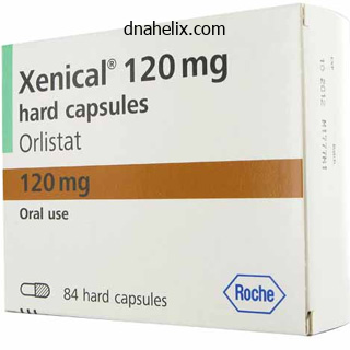
Xenical dosages: 120 mg, 60 mg
Xenical packs: 30 pills, 60 pills, 90 pills, 120 pills, 180 pills, 270 pills
Generic xenical 60 mg lineAnesthetic management is much like weight loss questions buy 60mg xenical fast delivery that for open procedures, besides that one-lung air flow is required (as against being desirable) for nearly all procedures. These procedures are complicated by the want to share the airway with the surgeon or pulmonologist; fortuitously, these procedures are sometimes brief. One of three techniques can then be used during rigid bronchoscopy: (1) apneic oxygenation using a small catheter positioned alongside the bronchoscope to insufflate oxygen (see above); (2) typical air flow through the side arm of a ventilating bronchoscope (when the proximal window of this instrument is opened for suctioning or biopsies, ventilation should be interrupted); or (3) jet air flow through an injector-type bronchoscope. Mediastinoscopy Mediastinoscopy, far more commonly employed up to now than at present, supplies entry to the mediastinal lymph nodes and is used to establish both the prognosis or the resectability of intrathoracic malignancies (see above). Mediastinoscopy is carried out under basic tracheal anesthesia with neuromuscular paralysis. A: the catheter is superior previous the obstruction, and the cuff is deflated when jet air flow is initiated. Jet ventilation may be continued with out interruption during resection and reanastomosis. Because the innominate artery could additionally be compressed in the course of the procedure, blood pressure must be measured in the left arm. Complications related to mediastinoscopy embrace (1) vagally mediated reflex bradycardia from compression of the trachea or the good vessels; (2) excessive hemorrhage; (3) cerebral ischemia from compression of the innominate artery (detected with a proper radial arterial line or pulse oximeter on the right hand); (4) pneumothorax (usually presents postoperatively); (5) air embolism (because of a 30� head elevation, the danger is biggest during spontaneous ventilation); (6) recurrent laryngeal nerve harm; and (7) phrenic nerve harm. Bronchoalveolar Lavage Bronchoalveolar lavage may be employed for sufferers with pulmonary alveolar proteinosis. In such patients, bronchoalveolar lavage may be indicated for extreme hypoxemia or worsening dyspnea. Unilateral bronchoalveolar lavage is performed beneath general anesthesia with a double-lumen bronchial tube. The cuffs on the tube ought to be properly positioned and should make a watertight seal to stop spillage of fluid into the opposite side. The process is often carried out in the supine position; though lavage with the lung in a dependent position helps to reduce contamination of the other lung, this position could cause severe ventilation/perfusion mismatch. Warm normal saline is infused into the lung to be treated and is drained by gravity At the end of the procedure, each lungs are nicely suctioned, and the double-lumen tracheal tube is replaced with a single-lumen tracheal tube. Preoperative Management Effective coordination between the organ-retrieval team and the transplant team minimizes graft ischemia time and avoids unnecessary prolongation of pretransplant anesthesia time. These procedures are performed on an emergency basis; due to this fact, patients could have little time to quick for surgical procedure. Administration of a clear antacid, an H2 blocker, or metoclopramide must be thought-about. Any premedication is normally administered solely within the operating room when the affected person is immediately attended and monitored. Immunosuppressants and antibiotics are additionally administered after induction and previous to surgical incision. Lung transplantation (as is true for all solid organ transplants) is proscribed by the availability of suitable organs, not by the availability of recipients. Intraoperative Management Monitoring Strict asepsis should be observed for invasive monitoring procedures. Central venous access could be accomplished solely after induction of anesthesia as a outcome of patients might not be succesful of lie flat whereas awake. Patients with a patent foramen ovale are susceptible to paradoxical embolism because of potentially high right atrial pressures. Transesophageal echocardiography is used to assess proper ventricular perform, integrity of the intraatrial septum, and pulmonary vein flow following anastomosis. Cystic fibrosis Bronchiectasis Obstructive Chronic obstructive pulmonary illness 1-Antitrypsin deficiency Pulmonary lymphangiomatosis Restrictive Idiopathic pulmonary fibrosis Primary pulmonary hypertension Induction & Maintenance of Anesthesia Induction with ketamine, etomidate, an opioid, or a combination of these agents is employed, avoiding precipitous drops in blood pressure. Inhalational agents are administered as tolerated for anesthesia and to provide a potential lung-protective effect. Hypercarbia and acidosis might result in pulmonary vasoconstriction and acute proper coronary heart failure, and hemodynamic assist with inotropes may be required for these sufferers. Drugs such as milrinone may be used for inotropic assist, and inhaled nitric oxide may be delivered to dilate the pulmonary vasculature. After the recipient lung is removed, the pulmonary artery, left atrial cuff (with the pulmonary veins), and bronchus of the donor lung are anastomosed. Flexible bronchoscopy is used to look at the bronchial suture line after its completion. Pulmonary vasodilators, inhaled nitric oxide, and inotropes (see earlier discussion) could additionally be necessary. Transesophageal echocardiography is helpful in identifying proper or left ventricular dysfunction, as nicely as in evaluating blood flow in the pulmonary vessels after transplantation. Transplantation disrupts the neural innervation, lymphatic drainage, and bronchial circulation of the transplanted lung. The respiratory pattern is unaffected, but the cough reflex is abolished below the carina. Loss of lymphatic drainage increases extravascular lung water and predisposes the transplanted lung to pulmonary edema. Loss of the bronchial circulation predisposes to ischemic breakdown of the bronchial suture line. Postoperative Management Patients are extubated after surgical procedure as soon as is feasible. A thoracic epidural catheter could also be employed for postoperative analgesia when coagulation studies are normal. The postoperative course could also be difficult by acute rejection, infections, and renal and hepatic dysfunction. Frequent bronchoscopy with transbronchial biopsies and lavage are essential to differentiate between rejection and infection. Nosocomial gram-negative bacteria, cytomegalovirus, Candida, Aspergillus, and Pneumocystis jiroveci are common pathogens. Other postoperative surgical issues embody damage to the phrenic, vagus, and left recurrent laryngeal nerves. Double-Lung or Heart�Lung Transplantation A "clamshell" transverse sternotomy can be used for double-lung transplantation. Posttransplantation Management After anastomosis of the donor organ or organs, eight ventilation to both lungs is resumed. Squamous cell carcinomas account for almost all of esophageal tumors; adenocarcinomas are much less frequent, whereas benign tumors (leiomyomas) are uncommon. After esophageal resection, the stomach is pulled up into the thorax, or the esophagus is functionally changed with a half of the colon (colonic interposition). Gastroesophageal reflux is handled surgically when the esophagitis is refractory to medical management or ends in complications such as stricture, recurrent pulmonary aspiration, or a Barrett esophagus (columnar epithelium). A number of antireflux operations may be performed (Nissen, Belsey, Hill, or Collis�Nissen) through thoracic or stomach approaches, typically laparoscopically. Achalasia and systemic sclerosis (scleroderma) account for almost all of surgical procedures performed for motility disorders. The former usually happens as an isolated discovering, whereas the latter is a part of a generalized collagen�vascular dysfunction. Cricopharyngeal muscle dysfunction can be related to a selection of neurogenic or myogenic issues and sometimes ends in a Zenker diverticulum.
Xenical 60 mg with mastercardPainful issues that usually reply to weight loss pills amphetamine buy xenical online pills sympathetic blocks include complicated regional ache syndrome, deafferentation syndromes due to nerve avulsion or amputations, and postherpetic neuralgia. However, the simplistic concept of heightened sympathetic activity resulting in vasoconstriction, edema, and hyperalgesia fails to account for the warm and erythematous part noticed in some sufferers. Psychological mechanisms or environmental elements are rarely the sole mechanisms for persistent ache but are generally seen together with different mechanisms (Table 47�6). The ache pathways mediating the afferent limb of this response are mentioned above. Sympathetic activation will increase efferent sympathetic tone to all viscera and releases catecholamines from the adrenal medulla. The hormonal response results from increased sym8 pathetic tone and from hypothalamicallymediated reflexes. Cardiovascular Effects Cardiovascular effects of acute ache often embody hypertension, tachycardia, enhanced myocardial irritability, and increased systemic vascular resistance. Cardiac output will increase in most traditional patients however could lower in patients with compromised ventricular perform. Because of the increase in myocardial oxygen demand, ache can worsen or precipitate myocardial ischemia. Respiratory Effects An increase in complete physique oxygen consumption and carbon dioxide manufacturing necessitates a concomitant enhance in minute ventilation. The latter increases the work of respiration, significantly in patients with underlying lung illness. Pain because of stomach or thoracic incisions further compromises pulmonary function due to guarding (splinting). Decreased motion of the chest wall reduces tidal quantity and functional residual capability, promoting atelectasis, intrapulmonary shunting, hypoxemia, and, much less generally, hypoventilation. Gastrointestinal and Urinary Effects Enhanced sympathetic tone will increase sphincter tone and reduces intestinal and urinary bladder motility, selling ileus and urinary retention. In addition, systemic opioids used to treat postoperative ache (and also administered as a component of the operative anesthetic) are a common reason for postoperative ileus and urinary retention. Endocrine Effects Stress increases release of catabolic hormones (catecholamines, cortisol, and glucagon) and inhibits release of anabolic hormones (insulin and testosterone). Patients develop a unfavorable nitrogen steadiness, carbohydrate intolerance, and increased lipolysis. The enhance in cortisol, renin, angiotensin, aldosterone, and antidiuretic hormone ends in sodium retention, water retention, and secondary enlargement of the extracellular space. Hematological Effects the neuroendocrine stress response to acute pain might increase platelet adhesiveness, scale back fibrinolysis, and promote a hypercoagulability state. Immune Effects the neuroendocrine stress response produces leukocytosis and may predispose patients to an infection. Worsening carbohydrate intolerance with sustained hyperglycemia also will increase the chance of an infection. Psychological Effects Anxiety and sleep disturbances are common reactions to acute ache. Some sufferers react with frustration and anger which could be directed at household, pals, and the medical staff. Systemic Responses to Chronic Pain the neuroendocrine stress response within the setting of persistent pain is mostly observed solely in sufferers with extreme recurring ache as a result of peripheral (nociceptive) mechanisms and in sufferers with outstanding central mechanisms corresponding to ache associated with paraplegia. Many patients also expertise important decreases or will increase in urge for food and psychological stress associated to social relationships. Evaluation of the Patient with Chronic Pain ought to include a number of key components. Information about location, onset, and high quality of ache, in addition to alleviating and exacerbating components, should be obtained, together with a ache historical past that features earlier therapies and modifications in signs over time. In addition to physical signs, chronic pain normally includes a psychological element that must be addressed. Information gathered in the course of the bodily examination might help distinguish ache location, type, and systemic sequelae, if any. All components are essential for a complete evaluation of the ache affected person prior to figuring out appropriate remedy options. It contains 20 units of descriptive words which are divided into four major groups: 10 sensory, 5 affective, 1 evaluative, and 4 miscellaneous. The affected person selects the sets that apply to his or her ache and circles the phrases in each set that finest describe the pain. It is often difficult to decide the relative contribution of despair to the struggling associated with ache. The Beck Depression Inventory is a useful check for identifying sufferers with main depression. Several exams have been developed to assess useful limitations or impairment (disability) and quality of life. Emotional problems are commonly associated with complaints of continual ache, and chronic ache often results in varying levels of psychological misery. Table 47�7 lists emotional disorders in which treatment must be primarily directed on the emotional dysfunction. Clear definitions are necessary, as pain could also be described in phrases of tissue destruction or bodily or emotional response. In the numerical scale, zero corresponds to no ache and 10 is intended to mirror the worst possible ache. The patient is requested to level to numerous facial expressions ranging from a smiling face (no pain) to a particularly sad one that expresses the worst attainable pain. The time between the onset of the stimulation and the onset of the muscle potential (latency) is a measurement of the quickest conducting motor fibers in the nerve. The amplitude of the recorded potential indicates the number of functional motor items, whereas its length displays the range of conduction velocities in the nerve. Conduction velocity could be obtained by stimulating the nerve from two factors and comparing the latencies. When a pure sensory nerve is evaluated, the nerve is stimulated while action potentials are recorded either proximally or distally (antidromic conduction). Nerve conduction studies distinguish between mononeuropathies (due to trauma, compression, or entrapment) and polyneuropathies. The latter includes acute or persistent neuropathies that are widespread and symmetrical (eg, associated to diabetes, alcohol abuse, malnutrition, toxins, or medication such as chemotherapeutic agents), or which might be focal however random (eg, mononeuropathy multiplex). Genetic factors and repetitive macrotrauma or microtrauma are likely concerned, and adjoining tenosynovitis is usually responsible. When a sensory nerve is involved, sufferers complain of ache and numbness in its distribution distal to the positioning of entrapment; often, a affected person might complain of ache referred proximal to the site of entrapment. Even entrapments of "pure" motor nerves can produce a vague ache which might be mediated by afferent fibers from muscles and joints. The diagnosis can often be confirmed radicular syndromes, neural trauma, and polyneuropathies.

Discount xenical 60mg on-lineLung resection could also be undertaken for progressive dyspnea or recurrent pneumothorax weight loss pills without exercise xenical 60 mg amex. The biggest risk of anesthesia is rupture of the air cavity during positive-pressure ventilation, leading to rigidity pneumothorax; the latter may occur on both aspect prior to thoracotomy or on the nonoperative side during the lung resection. Anesthesia for Tracheal Resection Preoperative Considerations Tracheal resection is most commonly carried out for tracheal stenosis, tumors, or, less commonly, congenital abnormalities. Tracheal stenosis may end up from penetrating or blunt trauma, in addition to tracheal intubation and tracheostomy. The dyspnea may be worse when the affected person is mendacity down, with progressive airway obstruction. Anesthetic Considerations Little premedication is given, as most sufferers presenting for tracheal resection have moderate to severe airway obstruction. Use of an anticholinergic agent to dry secretions is controversial because of the theoretical risk of inspissation. An inhalation induction (in 100 percent oxygen) is carried out in sufferers with severe obstruction. Laryngoscopy is carried out solely when the affected person is judged to be beneath deep anesthesia. Intravenous lidocaine (1�2 mg/kg) can deepen the anesthesia with out miserable respirations. The surgeon could then perform rigid bronchoscopy to evaluate and possibly dilate the lesion. Following bronchoscopy, the patient is intubated with a tracheal tube small enough to be handed distal to the obstruction every time possible. The surgeon divides the trachea in the neck and advances a sterile armored tube into the distal trachea, passing off a sterile connecting respiration circuit to the anesthesiologist for air flow in the course of the resection. Return of spontaneous air flow and early extubation on the end of the procedure are desirable. Surgical administration of low tracheal lesions requires a median sternotomy or proper posterior thoracotomy. Most procedures are carried out via three or extra small incisions within the chest, with the patient within the lateral decubitus place. This may result from obstruction, altered motility, or irregular sphincter function. In reality, most patients sometimes complain of dysphagia, heartburn, regurgitation, coughing, or wheezing when lying flat. Dyspnea on exertion may be prominent when continual aspiration leads to pulmonary fibrosis. Esophageal cancer patients normally have a history of cigarette smoking and alcohol consumption, so patients ought to be evaluated for coexisting persistent obstructive pulmonary 9 Regardless of the process, a standard anes- disease, coronary artery disease, and liver dysfunction. Patients with systemic sclerosis (scleroderma) ought to be evaluated for involvement of other organs, significantly the kidneys, heart, and lungs; Raynaud phenomena is also frequent. In patients with reflux, consideration must be given to administering a quantity of of the next preoperatively: metoclopramide, an H2-receptor blocker, sodium citrate, or a proton-pump inhibitor. The anesthesiologist may be requested to pass a large-diameter bougie into the esophagus as part of the surgical process; great warning should be exercised to assist avoid pharyngeal or esophageal injury. The former requires an higher stomach incision and a left cervical incision, whereas the latter requires posterolateral thoracotomy, an stomach incision, and, finally, a left cervical incision. During the transhiatal strategy to esophagectomy, substernal and diaphragmatic retractors can interfere with cardiac function. Colonic interposition entails forming a pedicle graft of the colon and passing it through the posterior mediastinum up to the neck to take the place of the esophagus. This process is lengthy, and upkeep of an sufficient blood stress, cardiac output, and hemoglobin concentration is necessary to ensure graft viability. Goal-directed fluid remedy utilizing hemodynamic measures (eg, stroke volume variation) may be helpful in perioperative fluid management of the esophagectomy patient. Lung protecting air flow and multimodal perioperative analgesia should be used postoperatively. Tracheal compression might produce dyspnea (proximal obstruction) or a nonproductive cough (distal obstruction). Asymptomatic compression can be frequent and may be evident only as tracheal deviation on physical or radiographic examinations. Flow�volume loops may also detect subtle airway obstruction and supply essential data regarding the situation and practical significance of the obstruction (see earlier discussion). Does the absence of any preoperative dyspnea make extreme intraoperative respiratory compromise much less likely Severe airway obstruction can happen following induction of anesthesia in these patients even in the absence of any preoperative signs. Moreover, lack of spontaneous ventilation can precipitate full airway obstruction. Superior vena cava syndrome is the outcome of progressive enlargement of a mediastinal mass and compression of mediastinal constructions, notably the vena cava. Lymphomas are most commonly responsible, but main pulmonary or mediastinal neoplasms also can produce the syndrome. Superior vena cava syndrome is usually associated with extreme airway obstruction and cardiovascular collapse on induction of general anesthesia. Caval compression produces venous engorgement and edema of the top, neck, and arms. Direct mechanical compression, in addition to mucosal edema, severely compromise airflow in the trachea. Most patients favor an upright posture, as recumbency worsens the airway obstruction. Cardiac output could additionally be severely depressed as a result of impeded venous return from the higher physique, direct mechanical compression of the heart, and (with malignancies) pericardial invasion. An echocardiogram is helpful in evaluating cardiac function and detecting pericardial fluid. Therefore, biopsy of a peripheral node (usually cervical or scalene) under local anesthesia is safest each time potential. Although establishing a analysis is of prime significance, the presence of significant airway compromise or the superior vena cava syndrome might dictate empiric remedy with corticosteroids previous to tissue analysis at surgery (cancer is the most common cause); preoperative radiation therapy or chemotherapy can also be thought of. The patient can usually safely endure surgery with general anesthesia once airway compromise and other manifestations of the superior vena cava syndrome are alleviated. How does the presence of airway obstruction and the superior vena cava syndrome influence administration of basic anesthesia The patient must be transported to the operating room in a semiupright position with supplemental oxygen. At least one large-bore intravenous catheter ought to be positioned in a decrease extremity, as venous drainage from the upper body may be unreliable. Airway administration: Difficulties with ventilation and intubation should be anticipated.
Purchase xenical with a visaA totally soaked "4 � 4" is mostly thought-about to hold 10 mL of blood weight loss log buy xenical pills in toronto, whereas a soaked "lap" may maintain 100 to 150 mL. More correct estimates are obtained if sponges and "laps" are weighed earlier than and after use, which is especially essential during pediatric procedures. Use of irrigating options complicates estimates, however their volume must be famous and an try made to compensate. Other Fluid Losses Many surgical procedures are related to obligatory losses of fluids other than blood. Such losses are due mainly to evaporation and inner redistribution of body fluids. Evaporative losses are most important with large wounds, particularly burns, and are proportional to the surface area uncovered and to the duration of the surgical process. Internal redistribution of fluids-often referred to as third-spacing-can trigger large fluid shifts and severe intravascular depletion in sufferers with peritonitis, burns, and similar conditions characterized by inflamed or infected tissue. Although estimates are difficult by occult bleeding into the wound or under the surgical drapes, accuracy is important to guide fluid therapy and transfusion. Shifting of intravascular fluid into the interstitial area (edema) is particularly important; protein-free fluid shift throughout an intact vascular barrier into the interstitial house is exacerbated by hypervolemia (water and sodium excess), and pathological alteration of the vascular barrier permits protein-rich fluid shift. Replacing Blood Loss Ideally, blood loss should be replaced with enough crystalloid or colloid options to keep normovolemia till the danger of anemia outweighs the risks of transfusion. At that time, further blood loss is changed with transfusion of red blood cells to preserve hemoglobin concentration (or hematocrit) at a suitable level. The level the place the benefits of transfusion outweigh its dangers should be considered on an individual foundation. Below a hemoglobin concentration of 7 g/dL, the resting cardiac output will increase to preserve a traditional oxygen supply. The transfusion point could be determined preoperatively from the hematocrit and by estimating 2 blood quantity (Table 51�5). Selection of the sort of intravenous resolution is decided by the surgical process and the anticipated blood loss. For minor procedures involving minimal or no blood loss, minimal or no fluid is commonly administered apart from for drug supply and for upkeep of intravenous line patency. The quantity of blood loss essential for the hematocrit to fall to 30% could be calculated as follows: 1. Degree of Tissue Trauma Minimal (eg, herniorrhaphy) Moderate (eg, open cholecystectomy) Severe (eg, open bowel resection) Additional Fluid Requirement 0�2 mL/kg 2�4 mL/kg 4�8 mL/kg four. Clinical pointers for transfusion generally used embody the following: (1) transfusing 1 unit of pink blood cells will enhance hemoglobin 1 g/dL and the hematocrit 2% to 3% in adults; and (2) a 10-mL/kg transfusion of purple blood cells will enhance hemoglobin concentration by three g/dL and the hematocrit by 10%. These extra fluid losses can be changed based on Table 51�6, primarily based on whether tissue trauma is minimal, average, or extreme. These values are only tips, and precise wants range considerably from affected person to affected person. Individuals often produce antibodies (alloantibodies) to the alleles they lack inside each system. Antibodies might occur spontaneously or in response to sensitization from a previous transfusion or pregnancy. Almost all individuals not having A or B antigen "naturally" produce antibodies, mainly immunoglobulin (Ig) M, towards these lacking antigens within the first 12 months of life. Approximately 85% of the white population and 92% of the black population has the D antigen, and people missing this antigen are called Rh-negative. Antibody screens are routinely accomplished on all donor blood and are frequently carried out for a potential recipient instead of a crossmatch (described next). Fortunately, with some exceptions (Kell, Kidd, Duffy, and Ss), alloantibodies against these antigens rarely trigger serious hemolytic reactions. Crossmatch A crossmatch mimics the transfusion: Donor red cells are mixed with recipient serum. Crossmatching, nonetheless, assures optimal security and detects the presence of less frequent antibodies not normally examined for in a screen. The probability of creating anti-D antibodies after a single publicity to the Rh antigen is 50% to 70%. Red blood cells, recent frozen plasma, and platelets are often transfused in a balanced ratio (1:1:1) in massive transfusion protocols and in trauma damage control resuscitation (see later discussion and Chapter 39). Nearly all models collected are separated into their part parts (ie, pink cells, platelets, and plasma), and whole blood items are hardly ever out there for transfusion in civilian practice. Red cells are usually saved at 1�C to 6�C however may be frozen in a hypertonic glycerol answer for up to 10 years. The latter method is usually reserved for storage of blood with uncommon phenotypes. The unit of platelets obtained generally contains 50 to 70 mL of plasma and may be saved at 20�C to 24�C for 5 days. One unit of blood yields about 200 mL of plasma, which is frozen for storage; as soon as thawed, it have to be transfused inside 24 h. Most platelets at the second are obtained from donors by apheresis, and a single platelet apheresis unit is equal to the amount of platelets derived from 6 to eight units of whole blood. The use of leukocyte-reduced (leukoreduction) blood products has been quickly adopted by many countries, including the United States, to find a way to decrease the risk of transfusion-related febrile reactions, infections, and immunosuppression. Surgical patients require quantity as well as purple cells, and crystalloid or colloid can be infused concurrently via a second intravenous line for quantity replacement. Blood for intraoperative transfusion ought to be warmed to 37�C during infusion, notably when more than 2 to 3 models will be transfused; failure to achieve this can lead to profound hypothermia. Granulocyte Transfusions Granulocyte transfusions, ready by leukapheresis, may be indicated in neutropenic patients with bacterial infections not responding to antibiotics. Transfused granulocytes have a really brief circulatory life span, so that day by day transfusions of 1010 granulocytes are usually required. Irradiation of those units decreases the incidence of graft-versus-host reactions, pulmonary endothelial damage, and different problems related to transfusion of leukocytes (see subsequent section), but might adversely have an effect on granulocyte function. Platelets Platelet transfusions must be given to sufferers with thrombocytopenia or dysfunctional platelets within the presence of bleeding. Prophylactic platelet transfusions are additionally indicated in sufferers with platelet counts under 10,000 to 20,000 � 109/L because of an increased risk of spontaneous hemorrhage. Platelet counts less than 50,000 � 109/L are related to increased blood loss during surgery, and such thrombocytopenic patients typically receive prophylactic platelet transfusions previous to surgical procedure or invasive procedures. Vaginal delivery and minor surgical procedures may be performed in sufferers with normal platelet function and counts greater than 50,000 � 109/L. Administration of a single unit of platelets could also be anticipated to enhance the platelet rely by 5000 to 10,000 � 109/L, and with administration of a platelet apheresis unit, by 30,000 to 60,000 � 109/L. Rh sensitization can occur in Rh-negative recipients as a result of the presence of a few purple cells in Rhpositive platelet items.
Quality xenical 120 mgA staged closure with a temporary Silastic "silo" may be necessary weight loss pills hydroxycut reviews cheap xenical 120mg with visa, adopted by a second process a few days later for complete closure. Persistent vomiting depletes potassium, chloride, hydrogen, and sodium ions, inflicting hypochloremic metabolic alkalosis. Initially, the kidney tries to compensate for the alkalosis by excreting sodium bicarbonate within the urine. Later, as hyponatremia and dehydration worsen, the kidneys should preserve sodium even on the expense of hydrogen ion excretion (paradoxic aciduria). Omphaloceles occur at the base of the umbilicus, have a hernia sac, and are sometimes associated with different congenital anomalies corresponding to trisomy 21, diaphragmatic hernia, and cardiac and bladder malformations. Antenatal prognosis by ultrasound may be followed by elective cesarean part at 38 weeks and quick surgical restore. Perioperative management focuses on stopping hypothermia, an infection, and dehydration. These issues are usually more critical in gastroschisis, as the protective hernial sac is absent. Anesthetic Considerations Surgery ought to be delayed till fluid and electrolyte abnormalities have been corrected. The abdomen ought to be emptied with a nasogastric or orogastric tube; the tube should be suctioned with the patient in the supine and lateral positions. Diagnosis often requires distinction radiography, and all contrast media have to be suctioned from the abdomen earlier than induction. Techniques for intubation and induction range, but in all instances the Anesthetic Considerations the abdomen is decompressed with a nasogastric tube earlier than induction. Experienced clinicians have variously advocated awake intubation, rapid sequence intravenous induction, and even cautious inhalation induction in chosen patients. These neonates could additionally be at increased danger for respiratory depression and hypoventilation within the recovery room because of persistent metabolic (measurable in arterial blood) or cerebrospinal fluid alkalosis (despite neutral arterial pH). Anesthetic Considerations Patients with croup are managed conservatively with oxygen and mist therapy. Indications for intubation include progressive intercostal retractions, apparent respiratory fatigue, and central cyanosis. Anesthetic administration of a overseas physique aspiration is challenging, particularly with supraglottic and glottic obstruction. Experts advocate cautious inhalational induction for a supraglottic object and delicate upper airway endoscopy to remove the item, safe the airway, or each. When the thing is subglottic, a rapid-sequence or inhalational induction is usually adopted by rigid bronchoscopy by the surgeon or endotracheal intubation and flexible bronchoscopy. Surgical preferences may vary based on the dimensions of the affected person and the nature and site of the international body. Children with impending airway obstruction from epiglottitis present within the operating room for definitive prognosis by laryngoscopy adopted by intubation. A preoperative lateral neck radiograph could present a attribute thumblike epiglottic shadow, which is very particular but typically absent. The radiograph can be useful in revealing other causes of obstruction, similar to overseas bodies. Rapid onset and progression of stridor, drooling, hoarseness, tachypnea, chest retractions, and a choice for the upright position are predictive of airway obstruction. Total obstruction can occur at any second, and preparations for a potential tracheostomy have to be made prior to induction of basic anesthesia. In most instances, an inhalational induction is carried out with the patient in the sitting position, utilizing a volatile anesthetic and oxygen. Oral intubation with an endotracheal tube one-half to one dimension smaller than traditional is tried as quickly as an enough depth of anesthesia is established. Foreign physique aspiration is usually encountered in kids aged 6 months to 5 years. Commonly aspirated objects include peanuts, cash, small batteries, screws, nails, tacks, and small items of toys. Onset is usually acute and the obstruction may be supraglottic, glottic, or subglottic. Stridor is outstanding with the primary two, whereas wheezing is more common with the latter. Acute epiglottitis is a bacterial an infection (most generally Haemophilus influenzae type B) classically affecting 2- to 6-year-old kids but additionally often showing in older kids and adults. It rapidly progresses from a sore throat to dysphagia and complete airway obstruction. The time period supraglottitis has been suggested because the inflammation sometimes includes all supraglottic constructions. Epiglottitis has increasingly turn into a disease of adults because of the widespread use of H influenzae vaccines in youngsters. If intubation is unimaginable, rigid bronchoscopy or emergency tracheostomy have to be carried out. Sleep apnea and up to date infection improve the chance of postoperative problems and may necessitate admission. Although these extremes of pathology are unusual, all youngsters undergoing tonsillectomy or adenoidectomy must be thought of to be at increased risk for perioperative airway problems. Causative organisms are normally bacterial and include pneumococcus, H influenzae, Streptococcus, and Mycoplasma pneumoniae. Myringotomy, a radial incision in the tympanic membrane, releases any fluid that has amassed in the middle ear. A historical past of airway obstruction or apnea suggests an inhalational induction with out paralysis till the flexibility to ventilate with constructive strain is established. Although deep extubation decreases the prospect of laryngospasm and will prevent blood clot dislodgment from coughing, an awake extubation is generally preferred to cut back the likelihood of aspiration. Postoperative vomiting is widespread and gastric suctioning is normally carried out prior to extubation. One must be alert within the recovery room for postoperative bleeding, signs of which may embrace restlessness, pallor, tachycardia, or hypotension. If reoperation is important to management bleeding, intravascular quantity must first be restored. Evacuation of stomach contents with a nasogastric tube is adopted by a rapid-sequence induction. Because of the potential for bleeding and Anesthetic Considerations these are typically very quick (10�15 min) outpatient procedures. Characteristic abnormalities of interest to the anesthesiologist embody a short neck, atlantooccipital instability, irregular dentition, psychological retardation, hypotonia, and a big tongue. Associated abnormalities embody congenital coronary heart disease in 40% of patients (particularly endocardial cushion defects and ventricular septal defect), subglottic stenosis, tracheoesophageal fistula, persistent pulmonary infections, and seizures.

Cheap xenical 60mg on-linePropoxyphene with and without acetaminophen (Darvocet and Darvon) was withdrawn from the U weight loss kit generic xenical 60 mg fast delivery. Parenteral Opioid Administration Intravenous, intraspinal (epidural or intrathecal), or transdermal routes of opioid administration could also be utilized when the affected person fails to adequately respond to, or is unable to tolerate, oral regimens. In sufferers with most cancers, adjunctive remedies similar to surgery, radiation, chemotherapy, hormonal therapy, and neurolysis may be useful. Intravenous Opioid Therapy Parenteral opioid therapy is usually finest accomplished by intermittent or continuous intravenous infusion, or each, but can be given subcutaneously. Spinal Opioid Therapy the use of intraspinal opioids is a superb different for patients acquiring poor aid with different analgesic techniques or who experience unacceptable side effects. Epidural and intrathecal opioids provide ache reduction with substantially lower total doses of opioid and fewer unwanted side effects. When compared with intermittent boluses, continuous intraspinal infusion methods reduce drug requirements, reduce unwanted facet effects, and decrease the likelihood of catheter occlusion. Myoclonic exercise could additionally be often observed with intrathecal morphine or hydromorphone. Epidural or intrathecal catheters could be positioned percutaneously or implanted to provide long-term efficient pain reduction. Epidural catheters can be attached to lightweight exterior pumps that might be worn by ambulatory patients. A momentary catheter should be inserted first to assess the potential efficacy of the method. Correct placement of the everlasting catheter should be confirmed utilizing fluoroscopy with contrast dye. With the affected person in proper lateral decubitus position, access to the intrathecal space and to the anterior stomach wall is optimized. After the posterior incision is made, a needle is superior via the incision into the intrathecal house, and a catheter is superior via the needle into the posterior intrathecal space. After the proximal catheter end is anchored, the distal end of the catheter is tunneled around the flank, beneath the costal margin to the anterolateral side of the belly wall. Implantable techniques are most acceptable for sufferers with a life expectancy of several months or longer, whereas tunneled epidural catheters are appropriate for patients anticipated to live only weeks. Formation of an inflammatory mass (granuloma) on the tip of the intrathecal catheter might happen and will reduce efficacy. The most regularly encountered drawback related to intrathecal opioids is tolerance, which is normally, however not all the time, a slowly growing phenomenon. The catheter connecting the pump to the intrathecal space is tunneled across the flank. Superficial infections can be decreased by the use of a silver-impregnated cuff close to the exit website. Other problems of spinal opioid therapy include epidural hematoma, which can become clinically apparent either instantly following catheter placement or several days later, and respiratory despair. Respiratory melancholy secondary to spinal opioid overdose can be handled by lowering the pump infusion price to its lowest setting and initiating a naloxone intravenous infusion. The efficacy and appropriateness of opioid therapy of persistent benign pain has increasingly come into query as nicely. Centers for Disease Control and Prevention printed tips for prescribing opioids for chronic pain to assist mitigate risks (see Guidelines at end of chapter). Botulinum Toxin (Botox) OnabotulinumtoxinA (Botox) injection has been increasingly utilized in the remedy of pain syndromes. This toxin blocks acetylcholine released at the synapse in motor nerve endings but not sensory nerve fibers. Proposed mechanisms of analgesia embrace improved native blood move, relief of muscle spasms, and release of muscular compression of nerve fibers D. A very massive quantity of fentanyl (10 mg) offers a big pressure for transdermal diffusion. Transdermal fentanyl patches can be found in 25, 50, 75, and 100 mcg/h sizes that present drug for two to 3 days. The major obstacle to fentanyl absorption through the pores and skin is the stratum corneum. Because the dermis acts as a secondary reservoir, fentanyl absorption continues for several hours after the patch is eliminated. Major disadvantages of the transdermal route are its gradual price of drug supply onset and the lack to quickly change dosage in response to changing opioid requirements. Blood fentanyl levels rise and attain a plateau in 12 to 40 h, providing common concentrations of 1, 1. Large interpatient variability results in precise supply charges ranging from 50 to 200 mcg/h. Transdermal fentanyl patches are sometimes diverted for illicit use, leading to numerous unintentional fatalities. Diagnostic & Therapeutic Blocks Local anesthetic nerve blocks are useful in delineating ache mechanisms, they usually play a major role within the management of sufferers with acute or continual pain. Pain aid following diagnostic nerve blockade often carries favorable prognostic implications for a subsequent therapeutic sequence of blocks. This approach can establish patients exhibiting a placebo response and those with psychogenic mechanisms. The efficacy of nerve blocks is due to interruption of afferent nociceptive exercise, which may be in addition to, or in combination with, blockade of afferent and efferent limbs of irregular reflex exercise involving sympathetic nerve fibers and skeletal muscle innervation. The pain reduction incessantly outlasts the identified pharmacological duration of the agent employed by hours or as a lot as several weeks. Local anesthetic solutions could also be infiltrated regionally or injected at specific peripheral nerve, somatic plexus, sympathetic ganglia, or nerve root websites. Ultrasound-Guided Procedures the usage of ultrasound in interventional ache drugs has increased dramatically over the past decade as a result of its utility in exactly visualizing vascular, neural, and different anatomic structures, its position as a substitute for the use of fluoroscopy and radiocontrast agents, and progressive improvements in know-how resulting in higher visual photographs and higher simplicity of use. Procedures that will benefit from ultrasound steering include set off point injections, nerve blocks, and joint injections. Fluoroscopy Fluoroscopy is highly effective for visualizing bony buildings and observing the spread of radiopaque distinction brokers. Live fluoroscopy with distinction agent could additionally be used to reduce the chance of intravascular injection of therapeutic brokers. Care should be taken to avoid excessive radiation dosage and to make use of appropriate radiation shielding, given the risks of ionizing radiation to the affected person and to the well being care staff in the fluoroscopy suite. An 8- to 10-cm 22-gauge needle is inserted roughly three cm lateral to the angle of the mouth on the degree of the higher second molar. The needle is then superior posteromedially and angled superiorly to deliver it into alignment with the pupil within the anterior aircraft and with the mid-zygomatic arch in the lateral plane. Without entering the mouth, the needle ought to pass between the mandibular ramus and the maxilla, and lateral to the pterygoid course of to enter the cranium via the foramen ovale. After a unfavorable aspiration for cerebrospinal fluid and blood, native anesthetic is injected. The nerve is definitely located and blocked with local anesthetic at the supraorbital notch, which is located on the supraorbital ridge above the pupil. The supratrochlear branch may additionally be blocked with native anesthetic at the superior medial corner of the orbital ridge.
Order xenical ukHyperosmolality with out hypernatremia could also be seen during marked hyperglycemia or following the buildup of abnormal osmotically energetic substances in plasma (see earlier discussion) weight loss pills ephedra buy xenical on line amex. In the latter two instances, plasma sodium concentration may very well lower as water is drawn from the intracellular to the extracellular compartment. For every 100 mg/dL increase in plasma glucose focus, plasma sodium decreases roughly 1. Hypernatremic sufferers may be hypovolemic, euvolemic, or hypervolemic (Table 49�4). However, hypernatremia is type of all the time the end result of both a relative loss of water in extra of sodium (hypotonic fluid loss) or the retention of enormous portions of sodium. Even when kidney concentrating ability is impaired, thirst is often highly efficient in preventing hypernatremia. Hypotonic losses may be renal (osmotic diuresis) or extrarenal (diarrhea or sweat). Urinary sodium concentration is usually larger than 20 mEq/L with renal losses and fewer than 10 mEq/L with extrarenal losses. Hypernatremia & Normal Total Body Sodium Content this group of sufferers generally manifests signs of water loss with out overt hypovolemia except the water loss is massive. Occasionally transient hypernatremia is noticed with movement of water into cells following exercise, seizures, or rhabdomyolysis. The commonest cause of hypernatremia in conscious patients with normal whole body sodium content material is diabetes insipidus. Plasma sodium concentration is determined according to the ratio of sodium and potassium to total body water. Rarely, important hypernatremia could additionally be encountered in patients with central nervous system issues. These sufferers appear to have "reset" osmoreceptors that function at the next baseline osmolality. Treatment is generally directed at the underlying illness and ensuring an enough fluid intake. Volume depletion by a thiazide diuretic can paradoxically lower urinary output by lowering water supply to collecting tubules. The analysis is suggested by a history of polydipsia, polyuria (often >6 L/d), and the absence of hyperglycemia. The absence of thirst in unconscious people results in marked water losses and might rapidly produce hypovolemia. The prognosis is confirmed by Hypernatremia & Increased Total Body Sodium Content this situation most commonly outcomes from the administration of enormous quantities of hypertonic saline solutions (3% NaCl or 7. Patients with main hyperaldosteronism and Cushing syndrome can also have elevations in serum sodium concentration along with indicators of elevated sodium retention. Clinical Manifestations of Hypernatremia Neurological manifestations predominate in sufferers with symptomatic hypernatremia, and restlessness, lethargy, and hyperreflexia can progress to seizures, coma, and in the end death. Symptoms correlate more intently with the rate of movement of water out of mind cells than with absolutely the degree of hypernatremia. Rapid decreases in mind quantity can rupture cerebral veins and end in focal intracerebral or subarachnoid hemorrhage. Seizures and severe neurological injury are frequent, notably in youngsters with acute hypernatremia when plasma [Na+] exceeds 158 mEq/L. After 24 to forty eight h, intracellular osmolality begins to rise as a outcome of will increase in intracellular inositol and amino acid concentrations, and brain intracellular water content slowly returns to normal. Treatment of Hypernatremia the remedy of hypernatremia is aimed at restoring plasma osmolality to normal and correcting the underlying trigger. Water deficits should usually be corrected over forty eight h, as rapid correction (or overcorrection) could cause cerebral edema. Hypernatremic sufferers with decreased whole body sodium ought to be given isotonic fluids to restore plasma volume to normal previous to therapy with a hypotonic solution. Hypernatremic patients with increased whole physique sodium should be handled with a loop diuretic along with intravenous 5% dextrose in water. Rapid correction of hypernatremia can end result in seizures, mind edema, everlasting neurological injury, and even demise. If one assumes that hypernatremia in this case represents water loss solely, then whole body osmoles are unchanged. Note that this technique ignores any coexisting isotonic fluid deficits, which if present ought to be changed with an isotonic solution. Anesthetic Considerations Hypernatremia has been demonstrated to increase the minimal alveolar concentration for inhalation anesthetics in animal studies, but its scientific significance is extra closely related to the associated fluid deficits. Hypovolemia accentuates any vasodilation or cardiac depression from anesthetic agents and predisposes to hypotension and hypoperfusion of tissues. Decreases in the volume of distribution for medication necessitate dose reductions for many intravenous agents, whereas decreases in cardiac output enhance the uptake of inhalation anesthetics. Routine measurement of plasma osmolality in hyponatremic sufferers quickly excludes pseudohyponatremia. Because of this tremendous reserve, hyponatremia is almost at all times the result of a defect in urinary diluting capability (urinary osmolality >100 mOsm/kg or particular gravity >1. Rare situations of hyponatremia with out an abnormality in renal diluting capacity (urinary osmolality <100 mOsm/kg) are usually attributed to major polydipsia or reset osmoreceptors; the latter two conditions may be differentiated by water restriction. Clinically, hyponatremia is finest categorized according to total body sodium content material (Table 49�6). Hyponatremia associated with transurethral resection of the prostate is mentioned in Chapter 32. Hyponatremia & Low Total Body Sodium Progressive losses of each sodium and water ultimately lead to extracellular volume depletion. Renal losses are mostly associated to thiazide diuretics and end in a urinary [Na+] greater than 20 mEq/L. Extrarenal losses are sometimes gastrointestinal and often are associated with a urinary [Na+] of lower than 10 mEq/L. A main exception to the latter is hyponatremia as a end result of vomiting, which might find yourself in a urinary [Na+] greater than 20 mEq/L. Edematous problems include congestive coronary heart failure, cirrhosis, kidney failure, and nephrotic syndrome. Hyponatremia in these settings outcomes from progressive impairment of renal free water excretion and usually parallels underlying disease severity. Proposed mechanisms for this dysfunction embrace extra secretion of natriuretic peptides and altered sympathetic stimulation to the kidney. Clinical Manifestations of Hyponatremia Symptoms of hyponatremia are primarily neurological and result from an increase in intracellular water. Their severity is generally related to the rapidity with which extracellular hypoosmolality develops.
Generic xenical 60 mg visaThe anhepatic section begins with the vascular occlusion of the inflow to the liver and ends with reperfusion weight loss pills jonah buy genuine xenical on-line. Some facilities utilize venovenous bypass to prevent congestion of the visceral organs, enhance venous return, and possibly shield kidney operate. In the neohepatic phase, two pathophysiological events may happen on opening the portal vein and allowing reperfusion of the graft. The first is a reperfusion syndrome brought on by the cold, acidotic, hyperkalemic solution which will comprise emboli and vasoactive substances being flushed from the graft immediately into the vena cava. This could trigger hypotension, proper coronary heart dysfunction, arrhythmias, and even cardiac arrest, and could also be preempted to some extent by the prophylactic administration of calcium chloride and sodium bicarbonate. This may outcome from impaired reperfusion because of severe endothelial dysfunction, and, in uncommon instances, could lead to major nonfunction of the graft. The use of reduced-size and residing donor grafts has increased the organ availability on this affected person population. Postoperative Management Patients who bear liver transplantation are often severely debilitated and malnourished and have multiorgan dysfunction. A close watch of graft operate have to be maintained, with a low threshold for checking hepatic artery patency and flow. Postoperative bleeding, biliary leaks, and vascular thromboses could require surgical reexploration. Living Donor Transplantation the usage of dwelling donors has elevated the pool of organs out there for transplantation. However, this procedure does expose wholesome people to morbidity and mortality risks. Adequate postoperative analgesia is required so that comfortable donor patients may be extubated at the end of the process. Complications of this surgery for the donor patient embrace transient hepatic dysfunction, wound an infection, postoperative bleeding, portal vein thrombosis, biliary leaks, and, hardly ever, death. An elevated incidence of perioperative brachial plexus harm has been reported in donor patients. Orthotopic liver transplantation is normally performed in sufferers with end-stage liver disease who begin to experience life-threatening complications, particularly when such issues become unresponsive to medical or nontransplant surgical procedure. Transplantation is also carried out in sufferers with fulminant hepatic failure (from viral hepatitis or a hepatotoxin) when survival with medical management alone is judged unlikely. The most common indications for liver transplantation in children, in order of lowering frequency, are biliary atresia, inborn errors of metabolism (usually 1-antitrypsin deficiency, Wilson illness, tyrosinemia, and Crigler�Najjar type I syndrome), and postnecrotic cirrhosis. The commonest indications in adults are postnecrotic (nonalcoholic) cirrhosis, primary biliary cirrhosis, and sclerosing cholangitis, and, less generally, major malignant tumors in the liver. One-year survival charges for liver transplantations exceed 80% to 85% in some facilities. The success of this process owes a lot to the use of cyclosporine and tacrolimus for immunosuppressant therapy. Cyclosporine is usually initially mixed with corticosteroids and other agents (eg, mycophenolate and azathioprine). Tacrolimus has proved efficient in cyclosporine-resistant rejection and is the popular alternative to cyclosporine as the primary immunosuppressant agent. Additional elements influencing the improvement in liver transplantation consequence embrace a larger understanding and experience with transplantation and improved evaluation and monitoring with echocardiography. These procedures can be divided into three phases: a dissection (preanhepatic) section, an anhepatic part, and a neohepatic section. Dissection (preanhepatic) phase: Through a hockey stick incision, the liver is dissected so that it remains connected only by the inferior vena cava and portal vein. Large belly varices may delay the period of, and improve the blood loss associated with, this section. Anhepatic section: Once the donor liver graft is prepared, the portal vein is clamped, followed by the inferior vena cava above and under the liver. The donor liver is then anastomosed to the supra- and infrahepatic inferior venae cavae and the portal vein. Revascularization and biliary reconstruction (neohepatic or postanhepatic) part: Following completion of the venous anastomoses, venous clamps are faraway from the cava, allowing venous blood to return to the center. Next, the portal vein is slowly opened, permitting blood to flush out preservation fluid and different substances accumulated in the liver during its ischemic time. This reperfusion might result in hypotension, arrhythmias, or cardiac arrest- a state of affairs termed the reperfusion syndrome. Potential issues embody multiorgan dysfunction brought on by cirrhosis, massive blood loss, hemodynamic instability from clamping and unclamping the inferior vena cava and portal vein, metabolic penalties of the anhepatic phase, and air embolism and hyperkalemia. Preoperative coagulation defects, thrombocytopenia, and previous stomach surgery greatly improve blood loss. Extensive venous collaterals between the portal and systemic venous circulations additionally contribute to increased bleeding from the abdominal wall. Potential problems of huge blood transfusion embrace hypothermia, coagulopathies, hyperkalemia, citrate intoxication (hypocalcemia), and the potential transmission of infectious agents. Blood salvaging strategies are useful in reducing donor pink blood cell transfusion. Several large-bore (14-gauge or larger) intravenous catheters must be placed above the diaphragm. Efforts to minimize the danger of hypothermia should embody the use of fluid warming and forced-air surface warming units. Urinary output ought to be monitored all through surgery by way of an indwelling urinary catheter. Laboratory measurements represent an essential part of intraoperative monitoring. Similarly, frequent measurements of arterial blood gases, serum electrolytes, serum ionized calcium, and serum glucose are necessary to detect and appropriately treat metabolic derangements. These latter modalities not solely assess overall clotting and platelet perform, but can also detect fibrinolysis. Most patients ought to be thought-about as having a "full stomach," usually due to marked abdominal distention or current upper gastrointestinal bleeding. The semiupright (back up) place throughout induction prevents speedy oxygen desaturation and facilitates ventilation until the abdomen is open. Anesthesia is usually maintained with a volatile agent (usually isoflurane or sevoflurane), and an intravenous opioid (usually fentanyl or sufentanil). The concentration of the volatile agent must be limited to lower than 1 minimal alveolar concentration in patients with extreme encephalopathy. Many patients are routinely transferred to the intensive care unit intubated and mechanically ventilated on the finish of the operative procedure, though quick postoperative extubation could additionally be thought of if the patient is comfortable, cooperative, physiologically secure, and never hemorrhaging significantly. Periodic calcium chloride administration (1 g) is necessary, however must be guided by ionized calcium concentration measurements to avoid hypercalcemia. Sodium bicarbonate remedy could additionally be necessary and will equally be guided by arterial blood fuel evaluation.
References - Phillips DM. JCAHO pain management standards are unveiled. JAMA. 2000;284:428-429.
- Irwin M, Daniels M, Weiner H. Immune and neuro-endocrine changes after bereavement. Psychiatric Clin North Am 1987;10:449-65.
- Hashizume M, Uchiyama Y, Horai N, et al. Tocilizumab, a humanized antiinterleukin- 6 receptor antibody, improved anemia in monkey arthritis by suppressing IL-6-induced hepcidin production. Rheumatol Int. 2010;30(7):917- 923.
- Israel, G.M., Bosniak, M.A. Follow-up CT of moderately complex cystic lesions of the kidney (Bosniak category IIF). AJR Am J Roentgenol 2003;181:627-633.
|

