|
Mr P Conaghan - Specialist Registrar
- John Radcliffe Hospital
- Oxford
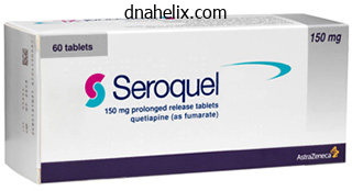
Seroquel dosages: 300 mg, 200 mg, 100 mg, 50 mg
Seroquel packs: 30 pills, 60 pills, 90 pills, 120 pills, 180 pills, 270 pills, 360 pills
Buy seroquel 100 mg lowest priceFurther analysis with radiologic imaging (commonly belly ultrasound medicine information cheap seroquel 200 mg mastercard, computed tomography, or magnetic resonance imaging) and endoscopy (commonly upper endoscopy or colonoscopy) is guided by the suspected underlying dysfunction. For aged, disabled, or marginally housed patients, it is very important elicit how they get hold of and put together their meals and the way they access toilet facilities. A basic survey ought to be performed to assess for indicators of weight loss (fat and muscle wasting), malnutrition (dry or skinny pores and skin, hair loss, edema, anasarca), and vitamin deficiencies (pellagra, scurvy). An oral examination looks for mucocutaneous candidiasis (which may reflect immunosuppression), ulcerations (which might replicate inflammatory bowel disease, vasculitis, viral infection, or vitamin deficiencies), and glossitis or angular cheilitis (which could reflect vitamin deficiencies). Examination of the lungs and cardiovascular system should focus on evidence of situations that may increase the risk of reasonable sedation within the occasion that endoscopy is required (respiratory insufficiency, heart failure) and of conditions that enhance the risk of intestinal ischemia (atrial fibrillation, valvular heart illness, peripheral vascular disease) (Chapter 134). Superficial or deep lots ought to be assessed for dimension, location, mobility, content (solid, liquid, or air), and the presence or absence of tenderness. Superficial masses embrace hernias, lymph nodes, subcutaneous abscesses, lipomas, and hematomas. Neoplasms (liver, gallbladder, pancreas, stomach, intestine, kidney), abscesses (appendicitis, diverticulitis, Crohn disease), or aortic aneurysms could represent deep belly plenty. Examination of the right higher quadrant ought to assess the liver dimension, contour, texture, and tenderness. Liver measurement is crudely estimated by percussion of the upper and decrease borders of liver dullness in the midclavicular line. Liver contour and tenderness are greatest assessed during held inspiration by deep palpation alongside the costal margin. The tip of an enlarged spleen may be palpated during inspiration if the examiner supports the left costal margin with the left hand while palpating under the costal margin with the best hand. Ascites must be suspected in a patient with a protuberant abdomen and bulging flanks. To display screen for ascites, percussion of the flanks must be carried out to assess the level of dullness. If the level of flank dullness seems to be increased, essentially the most sensitive take a look at for ascites is to examine for "shifting" dullness when the patient rolls from the supine to the lateral place. Digital Rectal and Pelvic Examinations the digital rectal examination is intrusive and uncomfortable and ought to be performed only when essential, such as in sufferers with perianal or rectal symptoms, incontinence, tough defecation, suspected inflammatory bowel illness, and acute belly pain. The perianal area ought to be visually inspected for rashes, soilage (suggesting incontinence or fistula), fistulas, fissures, pores and skin tags, exterior hemorrhoids, and prolapsed internal hemorrhoids (Chapter 136). After mild digital insertion, the anal canal should be assessed for resting tone and voluntary squeeze. The distal rectal vault must be swept circumferentially to palpate for mass lesions, tenderness, or fluctuance. Laboratory Studies Blood Tests Abdominal Examination the belly examination begins with a visual inspection of the abdomen and inguinal region for scars (due to prior surgical procedures or trauma), asymmetry (suggesting a mass or organomegaly), distention (due to weight problems, ascites, or intestinal ileus or obstruction), prominent periumbilical veins (suggesting portal hypertension), or hernias (umbilical, ventral, inguinal). The examination proceeds with auscultation adopted by percussion, and it ends with light and deep palpation. In sufferers without abdominal pain, auscultation of bowel sounds to assess intestinal motility has limited usefulness and could additionally be omitted. Percussion may be performed earlier than or in conjunction with mild and deep palpation. Initial cursory mild percussion throughout the higher, mid, and decrease stomach is helpful to denote areas of dullness and tympany as nicely as to elicit unanticipated areas of pain or tenderness before palpation. More in depth percussion provides limited however useful information about the scale of the liver and spleen, gastric or intestinal distention, bladder distention, and ascites (Chapters 137 and 144). Gentle, gentle palpation promotes belly relaxation and allows the detection of muscle resistance (guarding), stomach tenderness, and superficial lots of the stomach wall or abdomen. A low platelet count could also be attributable to portal hypertension with splenic sequestration. Abnormal liver check outcomes may be because of acute or chronic liver ailments, disorders of the pancreas or biliary tract, and medicines (Chapter 138). Serum amylase and lipase are obtained to screen for pancreatitis (Chapter 135) in patients with acute stomach pain. Increased ranges of inflammatory markers, similar to an elevated erythrocyte sedimentation price and C-reactive protein, are nonspecific however useful in the management of sufferers with inflammatory bowel illness (Chapter 132). Deficiencies within the fat-soluble nutritional vitamins (A, D, E, K) (Chapter 131) could mirror problems of malabsorption that lead to steatorrhea. Serum B12 could additionally be decreased in patients with autoimmune gastritis (pernicious anemia), gastric bypass surgery, or malabsorption due to small bowel bacterial overgrowth or illness of the terminal ileum. In patients with acute diarrhea, assessment of fecal leukocytes or tradition of widespread pathogens is routine, and in selected patients, testing for parasites (Giardia, Entamoeba histolytica), Clostridium difficile, Escherichia coli O157:H7, or other particular organisms could additionally be warranted. In many medical settings, commercially obtainable molecular diagnostic tests can screen patients with acute diarrhea for a defined panel of micro organism, viruses, and parasites, and supply outcomes inside 1 to 5 hours. Esophageal manometry and esophageal pH and impedance monitoring may be helpful for the analysis of heartburn, reflux, and different esophageal symptoms (Chapter 129). Anorectal manometry may be helpful in some patients with fecal incontinence and defecatory dysfunction (Chapter 136). Thereafter, the emphasis should swap from finding a "cause" of the symptoms to implementing profitable coping and adaptive behaviors. Abdominal ache, which is a frequent grievance amongst outpatients in the office setting and emergency department, may be benign and self-limited or the presenting symptom of extreme, life-threatening illness. By distinction, most patients with extreme acute abdominal ache require a radical however emergent analysis, which can quickly reveal an acute surgical illness (Chapter 133). Stimulation of hole stomach viscera is mediated by splanchnic afferent fibers within the muscle wall, visceral peritoneum, and mesentery that are delicate to distention and contraction. Visceral afferent nerves are loosely organized, innervate several organs, and enter the spinal twine at several levels. Thus, visceral pain is obscure or dull in character and diffuse; patients making an attempt to localize the ache usually move their complete hand over the upper, middle, or decrease stomach. Most visceral ache is regular, however cramping, intermittent ache or "colic" results from peristaltic contractions attributable to partial or full obstruction of the small intestine, ureter, or uterine tubes. In distinction to visceral innervation, a dense network of nerve fibers that observe a spinal T6 to L1 somatic distribution innervates the parietal peritoneum. Pain fibers of the parietal peritoneum are stimulated by stretch or distention of the stomach cavity or retroperitoneum; direct irritation from infection, pus, or secretions. Parietal pain is sharp, well characterized, and localized by the patient to a exact location on the stomach, usually by pointing with one finger. Severe abdominal ache that begins abruptly throughout seconds to minutes indicates a catastrophic event, similar to esophageal rupture, perforated peptic ulcer or viscus, ruptured ectopic being pregnant, ruptured aortic aneurysm, acute mesenteric ischemia, or myocardial infarction. Pain that progresses inside 1 to 2 hours is according to a rapidly progressive inflammatory dysfunction. The character of the ache provides necessary information about whether or not the signs are as a end result of visceral stimulation or parietal stimulation (peritonitis). Patients with peritonitis might report severe localized pain or irritation with activities or maneuvers that stretch or transfer the parietal peritoneum, corresponding to walking, moving in bed, and coughing; as a result, they have an inclination to lie quietly to avoid painful stimulation.
Seroquel 50mg low priceMalignant ulcers are characteristically irregular in shape with heaped borders symptoms food poisoning cheap seroquel online amex, but they also may be flat or depressed. Current highresolution and magnification endoscopes permit visualization of the altered mucosal structure surrounding an ulcer, including modifications in the microvascular sample. Primary gastric adenocarcinomas typically occur in mucosal areas with atrophic and intestinal metaplastic adjustments. For a particular analysis of malignancy, a quantity of biopsy specimens are wanted, normally from the ulcer margins. A few gastroduodenal ulcers are attributable to systemic inflammatory illnesses, particularly, Crohn disease (Chapter 132). Patients with Crohn illness affecting the proximal gastrointestinal tract usually have multiple ulcers characterised by irregular longitudinal shapes. The demonstration of ulcerative irritation elsewhere in the digestive tract, particularly within the terminal ileum and colon, strongly supports the diagnosis of Crohn disease, as do noncaseating granulomas on biopsy specimens. Other inflammatory disorders that may cause gastritis or gastroduodenal ulcers include various types of vasculitis affecting the mesenteric system, specifically Beh�et illness (Chapter 254), Henoch-Sch�nlein purpura (Chapter 254), Takayasu arteritis (Chapters 69 and 254), polyarteritis nodosa (Chapter 254), systemic lupus erythematosus (Chapter 250), Churg-Strauss syndrome (Chapter 254), and granulomatosis with polyangiitis (Chapter 254). Lymphocytic gastroduodenitis, which is strongly related to celiac illness (Chapter 131), might lead to duodenal ulceration and subsequent stenotic web formation. Ulcer disease additionally might happen in sufferers with polycythemia vera (Chapter 157), possibly in relation to lowered mucosal blood move. Vasculitis underlying ulcer disease must be thought-about in sufferers with chronic or recurrent ulceration in whom other causes have been excluded. Lymphocytic phlebitis, which is a uncommon vasculitic inflammatory disorder that affects the mesenteric veins, may cause gastric ulcers. Systemic amyloidosis (Chapter 179) affecting the abdomen wall may lead to gastric ulcers. Rare cases of duodenal ulceration have been described within the presence of annular pancreas or congenital bands obstructing the descending duodenum. The most essential hypergastrinemic disorder is Zollinger-Ellison syndrome (Chapter 219), a condition of marked hyperacidity resulting in extreme peptic ulcer illness attributable to a gastrin-producing endocrine tumor. These sufferers usually have a number of bulbar and postbulbar duodenal ulcers which might be immune to conventional acid suppressive therapy. The prognosis could be confirmed by the presence of a excessive fasting serum gastrin level (often but not all the time 10-fold elevated and >1000 pg/mL). Similar gastrin levels are generally seen in patients handled for chronic ulcer disease with high-dose proton pump inhibitors. For clarification, secretin testing may be performed: in patients with Zollinger-Ellison syndrome, the injection of secretin (1 U/ kg) increases serum gastrin ranges by greater than 50%, or a hundred and twenty pg/mL or higher, in these with fasting gastrin ranges less than 10-fold above regular. In some sufferers, Zollinger-Ellison syndrome occurs as a half of the a quantity of endocrine neoplasia syndrome (Chapter 218), notably in affiliation with hyperparathyroidism. Other hypergastrinemic hyperacidity syndromes are the retained gastric antrum syndrome (see later) and antral G-cell hyperfunction. Pallor of the mucosa, in preserving with decreased mucosal blood circulate, could also be famous at endoscopy. Upper mesenteric ischemia is usually associated with upper abdominal ache, which can be elicited by a meal or by bodily exercise. These signs might cause sufferers to lower their meals intake, leading to weight reduction before their clinical presentation. Alcohol Short-term heavy alcohol use or long-term reasonable to heavy alcohol use can result in indicators of acute and chronic gastritis. No proof signifies that this kind of gastritis is associated with a major threat for peptic ulceration, though alcohol use will increase the chance for bleeding in patients with peptic ulcer illness. Stress Ulcers Patients with extreme medical conditions, such as main trauma, sepsis, extensive burns, head harm, or multiorgan failure, can develop stress ulcers in the abdomen or duodenum. Major risk components for stress ulceration in severely unwell sufferers include mechanical ventilation, coagulopathy, and hypotension, however factors similar to hepatic and renal failure and the utilization of ulcerogenic drugs could contribute. Ulcers related to head harm are generally identified as Cushing ulcers, and ulcers associated with extensive burns are known as Curling ulcers. Stress ulcers had been once frequent in sufferers in intensive care units, however enhancements in general administration, including respiratory and hemodynamic care, acid inhibition, and emphasis on enough feeding, have lowered the incidence of these ulcers, which presently have an effect on 1 to 2% of those sufferers. Similar to the hypergastrinemic syndromes, persistent elevation of histamine can lead to hyperacidity because of the chronic stimulation of parietal cells. Systemic mastocytosis (Chapter 240) is characterized by a proliferation of mast cells within the bone marrow, pores and skin, liver, spleen, and gastrointestinal tract, usually associated with each spontaneous and trigger-induced. Patients with systemic mastocytosis often have gastrointestinal symptoms, including pain, diarrhea, and blood loss. Histamine hypersecretion leading to peptic ulcer disease can also occur in continual myelogenous leukemia (Chapter 175) with basophilia. Other Drugs Other Factors Cameron Ulcer Patients with giant hiatal hernias (Chapter 129) might current with proximal gastric ulcers, termed Cameron ulcers, at the degree of the hiatus, where the stomach is compressed. During higher gastrointestinal endoscopy, patients with giant hiatal hernias and iron deficiency anemia must be rigorously examined in regular and retroverted positions for the presence of Cameron ulcers. Ischemia and continual inflammation specifically ensuing from biliary reflux could cause such ulcers. Anastomotic ulcers after bariatric gastric surgery seem to be associated to local ischemia. Because it then lacks publicity to acid and is thus not physiologically downregulated, it continues to secrete gastrin regardless of regular and even excessive acid ranges. Oral bisphosphonates, used extensively for osteoporosis (Chapter 230), may induce gastric erosions and ulcerations in an estimated three to 10% of treated patients. Although corticosteroid treatment may be sophisticated by peptic ulcer illness, the relative threat is just slightly increased, except in sufferers with critical comorbid illnesses, using long-term or high-dose therapy, or with prior ulcers. Similarly, the use of aldosterone antagonists can be associated with an elevated threat for peptic ulcer and ulcer bleeding, probably associated to impaired mucosal therapeutic. Persons who use amphetamines and crack cocaine (Chapter 31) regularly develop ulcer disease, typically with perforation, presumably as a outcome of vascular insufficiency. Chemotherapy, notably when given selectively as a high-dose intra-arterial infusion within the celiac system, could be sophisticated by ulcer disease. Patients on anticoagulant therapy and people with other coagulopathies may not often develop intramural hematoma of the gastrointestinal tract. Radiation remedy of the upper stomach is typically complicated by persistent ischemic ulceration, particularly as a late complication. Indeed, a silent ulcer may be recognized only when it presents abruptly with a complication, mostly hemorrhage or perforation, or it might be discovered incidentally when a diagnostic take a look at is performed for other causes. Nevertheless, the standard presentation of acid peptic illness is recurrent episodes of pain. The ache is nearly invariably positioned within the epigastrium and will radiate to the again or, less commonly, to the thorax or different areas of the stomach (see Table 130-2). Some patients describe the pain as burning or piercing, whereas others describe it as an uncomfortable feeling of vacancy of the abdomen, referred to as painful hunger.
Seroquel 200mg amexA2 Replacing iron by intravenous route is proposed to forestall and deal with acute blood loss after major surgical procedure as a transfusion-sparing technique medicine gif purchase seroquel amex. Mild to reasonable infusion reactions, such as pruritus, urticaria, skin flushing, myalgia, and again pain, are common however simply controlled. Contraindications to intravenous iron are concomitant infections, severe atopy, and previous severe hypersensitivity reactions. Long-term unwanted facet effects in case of iron accumulation and susceptibility to infections stay unexplored, and the total quantity of iron that can be safely administered, particularly in sufferers with inflammatory circumstances, stays to be defined. Recommendations can be found for the administration of the rare atypical microcytic anemias because of genetic disorders of iron metabolism and heme synthesis. Sixty and 30 mg elemental oral iron every day or on alternate days are suggested in adults and children respectively. Routine prophylaxis with oral iron carries the risk of exacerbating malaria infections in endemic African areas where iron administration in affected children must be always accompanied by malaria remedy. Adequate treatment with oral or intravenous iron controls iron deficiency and iron deficiency anemia, and if the etiologic cause is reversible, a single cycle of remedy is enough to restore hemoglobin and ferritin levels. Relapse of iron deficiency after therapy often indicates persistence of the cause, which must be intensively investigated. Most mechanisms are adaptive: leukocytosis is a defense against pathogens, and hepcidin-induced hypoferremia impairs extracellular microorganism progress. High serum ferritin on account of inflammation and macrophage iron retention, related to lownormal iron and whole iron binding capability (or transferrin levels), and decreased % transferrin saturation recommend inflammation. Elevated C-reactive protein related to excessive ferritin levels suggests inflammation. Soluble transferrin receptor (sTfr), elevated in iron deficiency or when erythropoiesis is expanded, could help to differentiate the anemia of irritation and iron deficiency. In general, an increased sTfr is found in iron deficiency, with or with out coexisting inflammation. The sTfr/log ferritin index is taken into account one of the best indicator for discriminating anemia of irritation (ratio of about 1) from iron deficiency in anemia of inflammation (ratio of more than 2) (Table 150-4). In apply, the prognosis of iron deficiency in inflammatory circumstances relies on an arbitrary cutoff of ferritin (<100 g/L) or percent saturation of transferrin (<20%) in case the ferritin is greater than 100 g/L (usually between a hundred and 300). The anemia of irritation is the second most frequent anemia worldwide and the first amongst hospitalized sufferers. Acute types of anemia of irritation occur in intensive care models, secondary to sepsis, in depth burns, or polytrauma. The anemia of inflammation is related to elevated manufacturing of proinflammatory cytokines, upregulation of hepcidin, and deregulation of iron homeostasis. Pathogenic mechanisms are a quantity of, including relative erythropoietin deficiency, blunted erythropoietic response, macrophage iron sequestration, iron-restricted erythropoiesis, and shortened erythrocyte survival. Anemia in each hematologic malignancies (lymphoma and myeloma) and strong tumors (ovarian and colon cancer) is much more complex; inflammation in cancer is often current together with blood loss or chemotherapy-induced bone marrow toxicity, malnutrition, or concomitant infections. The anemia of irritation might coexist with absolute iron deficiency because of blood loss, as may incessantly happen in inflammatory bowel illnesses and colon cancer. In inflammatory bowel disease, intravenous iron ought to be preferred in case of illness flares as a outcome of oral iron is less tolerated and should even trigger further mucosal damage. They are used in selected patients with myelodysplastic syndromes and in most cancers patients present process chemotherapy. In this functional iron deficiency, intravenous iron is more practical than oral iron. Blood transfusions are indicated only to alleviate anemia-related signs but must be prevented as chronic therapy due to transfusion-associated risks. Sideroblastic Anemias the term sideroblastic anemia encompasses both congenital and bought anemias in which disruption of erythroblast iron utilization causes ineffective erythropoiesis and variable levels of systemic iron overload. A hallmark of sideroblastic anemia is the presence of bone marrow ring sideroblasts, erythroblasts with coarse blue granules at Perls staining, as a result of pathologic iron deposits in mitochondria. Congenital forms are inherited issues of mitochondria that could be nonsyndromic or syndromic, according to whether the mutation additionally impacts nonhematologic tissues. Acquired forms are clonal myelodysplastic (Chapter 172) or myelodysplastic-myeloproliferative issues (Chapter 157). Usually, the acquired sideroblastic anemias which might be usually associated with myelodysplasia (Chapter 172) have macrocytic pink cells. The mechanism of mitochondrial iron accumulation may be due to heme synthesis blockade and/or pyridoxal sulfate antagonism. If the mutation is unknown, an try with vitamin B6 (100 to 200 mg every day for two to three months) must be tried. Responsive sufferers ought to remain on low-dose pyridoxine (10 to 50 mg/day) lifelong. In the latter case, microcytosis helps the differential diagnosis of the acquired varieties. The few presenting with anemia have skewed chromosome X inactivation in hematopoietic lineage and are usually identified in maturity. The kind of mutation may be relevant for remedy: patients with mutations within or close to the cofactor (5-pyridoxal phosphate) binding web site respond to pyridoxine treatment; the others are pyridoxine refractory. The pathogenic lesions in all types are decreased erythroid iron utilization for heme synthesis, mitochondrial iron deposits, oxidative harm, and cell death. Because of the coordinated regulation of heme and globin, low heme reduces globin translation explaining microcytosis and hypochromia. All hereditary forms normally present more than 15% ringed sideroblasts where the iron deposition covers multiple third of the nucleus rim. Females with X-linked types and carriers of non-sense mutations might have normocytic or macrocytic erythrocytes. The diagnosis of hereditary sideroblastic anemias requires genetic testing with the identification of the mutation within the corresponding gene. Iron chelation should be started (usually when ferritin ranges are >1000 g/L) to keep away from iron toxicity. Iron chelation must be tried in iron-loaded patients as a end result of in a number of circumstances anemia is partially corrected by iron depletion. Effect of ferric carboxymaltose on exercise capability in patients with persistent coronary heart failure and iron deficiency. Effects of ferric carboxymaltose on hospitalisations and mortality charges in iron-deficient heart failure sufferers: an individual affected person knowledge meta-analysis. Guidelines on the analysis and treatment of iron deficiency throughout indications: a systematic review. Oral iron dietary supplements improve hepcidin and decrease iron absorption from every day or twice-daily doses in iron-depleted younger ladies. Intravenous iron remedy in patients with chronic kidney disease: recent proof and future instructions. International consensus assertion on the peri-operative administration of anaemia and iron deficiency.
Generic seroquel 50mg free shippingIt could additionally be a greater check of the integrity of systemic hemostasis than any laboratory measurement can present treatment quotes order seroquel in india. In a patient with a historical past of excessive or unexplained bleeding, the initial aim is to decide whether the cause is a systemic coagulopathy or localized anatomic or mechanical downside with a blood vessel. This scenario is encountered most regularly in sufferers with extreme postoperative bleeding, which could be due to both local surgical trauma or a coagulation abnormality, or each. A historical past of prior bleeding suggests a coagulopathy, as does the discovering of bleeding from multiple websites. Even diffuse bleeding may come up from anatomic rather than hemostatic abnormalities. An example of that is recurrent mucosal hemorrhage in patients with hereditary hemorrhagic telangiectasia (Chapter 164). Conversely, a single episode of bleeding from an isolated site may be the preliminary manifestation of a coagulopathy. The historical past must include a survey of coexisting systemic ailments and drug ingestions that would have an result on hemostasis. Renal failure and the myeloproliferative neoplasms are related to impaired platelet�vessel wall interactions and qualitative platelet abnormalities, connective tissue illnesses and lymphomas are associated with thrombocytopenia, and liver illness causes a posh coagulopathy (Chapter 166). Other drugs, corresponding to antibiotics, additionally could also be associated with a bleeding tendency by causing abnormal platelet perform or thrombocytopenia. Mild bleeding events are generally reported by patients with and with out subsequently laboratory-documented bleeding problems, generally making it tough for hematologists to outline a "significant bleeding historical past. More precise quantification of bleeding symptoms is being tried by using "bleeding rating" devices such as the Vicenza bleeding rating to assist discriminate, in conjunction with laboratory testing, between healthy topics and people with mild bleeding disorders. Patterns of medical bleeding, as revealed by the historical past and bodily examination, could additionally be characteristic of certain forms of coagulopathy (Table 162-1). These might contain petechiae, which are pinpoint cutaneous hemorrhages that seem significantly over dependent extremities (characteristic of severe thrombocytopenia), ecchymoses (common bruises), purpura, gastrointestinal and genitourinary tract bleeding, epistaxis, and hemoptysis. In most of these issues, bleeding tends to occur spontaneously or instantly after trauma. In distinction, patients with inherited or acquired coagulation factor deficiencies, similar to hemophilia, or these on anticoagulant therapy are probably to bleed from deeper tissue websites. Pseudothrombocytopenia is indicated by the finding of platelet clumps on the peripheral smear, and the prognosis is supported by the finding of simultaneously normal platelet counts in blood samples obtained by finger stick, in tubes containing different anticoagulants, or in a tube maintained at 37� C before platelet counting. Examination of the blood smear can also reveal clues to the cause for real thrombocytopenia, such as fragmented pink blood cells in thrombotic thrombocytopenic purpura. The bleeding time was a widely used scientific screening take a look at for disorders of platelet�vessel wall interactions. It measures the time to cessation of bleeding after a standardized incision over the volar aspect of the forearm. However, the take a look at is susceptible to problems related to high quality management, reproducibility, sensitivity, and specificity. As an extra substitute for the bleeding time, especially when a practical (qualitative) abnormality of platelets is suspected by characteristic mucocutaneous bleeding or bruising, a global assay of platelet operate may be appended to the panel of screening exams. With a few notable exceptions, regular results for all these screening checks of hemostasis primarily exclude any clinically vital systemic coagulopathy. A to C, Algorithms for clinical and laboratory approach to the prognosis of a patient with a suspected systemic bleeding disorder (coagulopathy). This test supplies a world assay of coagulation and fibrinolysis using pointof-care know-how. It is a complete blood clotting test by which a small aliquot of blood is rotated in a cuvette, and the power, elasticity, and dissolution of the forming clot are measured with a torsion wire or by optical detection. A1 Its influence on producing other favorable clinical end factors stays open to study. Approach to evaluating patients with prolonged prothrombin time (pt) or activated partial thromboplastin time (aptt). If correction happens with the inhibitor display screen, particular person coagulation issue levels must be assayed for a specific deficiency state. These tests measure primarily the coagulation arm of hemostasis and have been shown to be promising in evaluating thrombophilias in guiding antithrombotic therapy. The exams are limited right now by their lack of validation as a clinical device. It is essential to view the clinical setting, history, physical examination, and screening laboratory exams as complementary sides of the strategy to sufferers with suspected coagulopathies. Laboratory testing and probably further specialised tests of coagulation are indicated for sufferers whose bleeding histories are suspicious for a hemostatic abnormality. Patients usually have deep vein thrombosis of the decrease extremities or pulmonary embolism, however different unusual websites of venous thrombosis could also be concerned. Arterial thrombosis that occurs prematurely or within the absence of apparent danger components ought to trigger a special line of investigation, possibly including evaluation for vasculitis, myeloproliferative neoplasms, hyperhomocysteinemia, antiphospholipid syndrome, and potential sources of systemic embolization. The major or hereditary hypercoagulable states (see Table 73-1) end result from specific mutations or polymorphisms that result in decreased levels of physiologic antithrombotic proteins or elevated levels of procoagulant proteins. In contrast, the secondary or acquired hypercoagulable states are a heterogeneous group of problems that predispose to thrombosis by complicated mechanisms. Certain clinical characteristics suggest the presence of an inherited hypercoagulable state (Table 162-3). Patients with recurrent thrombosis should be examined for these problems and, in most cases, dedicated to lifelong prophylactic anticoagulation. Acute thrombosis itself can cause transient decreases within the ranges of antithrombin, protein C, and protein S. This association is increased additional in sufferers with recurrent and unprovoked thrombosis. There are conflicting opinions on whether or not an evaluation for occult malignancy in these sufferers have to be exhaustive. Most advocate that analysis could be restricted to an intensive historical past, bodily examination, routine full blood cell count and chemistries, take a look at of fecal occult blood, urinalysis, mammogram (in women), and chest radiograph, with additional testing guided by any abnormalities found on this preliminary analysis. In addition to classic deep vein thrombosis and pulmonary embolism, sure attribute kinds of thrombosis could present important clues to the cause and trigger a extra directed analysis. Migratory, superficial thrombophlebitis (Trousseau syndrome) or nonbacterial thrombotic endocarditis strongly suggests the presence of an occult malignancy (Chapter 73). Hepatic vein thrombosis (Budd-Chiari syndrome; Chapter 134) or portal vein thrombosis may point out a myeloproliferative neoplasm (Chapter 157) or paroxysmal nocturnal hemoglobinuria (Chapter 151). Extensive inferior vena cava thrombosis might occur with renal cell carcinoma (Chapter 187). Warfarin-induced pores and skin necrosis strongly suggests underlying protein C or protein S deficiency.
Buy seroquel 50 mg amexThe mobile apoptosis causes intramedullary hemolysis or "ineffective erythropoiesis medicine ball workouts 200mg seroquel. A, oval macrocytes and fragmented purple blood cells are present in a smear from a patient with severe cobalamin deficiency and thalassemia trait, demonstrating an extreme vary in dimension and shape of the red blood cells. C, a gaggle of megaloblastic pink cell precursors all with an open lacy chromatin sample is proven from a bone marrow aspirate. D, the bone marrow biopsy exhibits hypercellularity with a predominance of huge erythroblasts and lots of mitotic figures, all potentially confused with acute leukemia. The demyelinating lesion, often termed "subacute mixed degeneration," begins with signs of symmetric paresthesia, normally within the lower extremities but generally involving the hands first. There is early impairment of proprioception ending with loss of vibration and position sense manifesting as ataxic gait. As the severity of the spinal lesions will increase weak spot develops, with spasticity and hyperreflexia, and finally sufferers may develop a segmental cutaneous sensory stage and even paraplegia. Altered mental standing often happens particularly with irritability, delusions, paranoia, lability, even mania referred to as "megaloblastic insanity" prior to now. Infants present with unique abnormalities, particularly hypotonia, irritability, lethargy, failure to thrive, and occasionally motion disorders. The underlying biochemical lesions causing the neurologic disease are nonetheless unknown although changes in lipids, development factors, cytokines, or methylation standing of proteins have all been studied. There is a powerful inverse relationship between the severity of the neurologic abnormalities and the hematologic abnormalities, which has been demonstrated in a big series of patients with pernicious anemia and recognized for no less than one hundred years within the literature. There is also an inverse relationship between the length of neurologic abnormalities prior to prognosis and the completeness of correction after enough cobalamin substitute, so early analysis is imperative. Further dialogue of the neurologic abnormalities of megaloblastic anemia and also alcoholism seems in Chapter 388. Cytogenetic research may present chromosomal fragmentation and in some cases even clonal abnormalities which appropriate with vitamin alternative and resolution of megaloblastic anemia. There is indirect hyperbilirubinemia, low serum haptoglobin, and generally excessive values for lactate dehydrogenase due to the intramedullary hemolysis. The presence of hemolysis markers and fragmented cells current on peripheral blood smear with normal instead of low reticulocyte counts will recommend a hemolytic anemia to the unwary clinician, as the literature well documents. To complicate the situation additional, there are stories of previously undiagnosed defects of cobalamin metabolism corresponding to cobalamin-C mutation presenting with hemolytic-uremic syndrome. Other tissues which have speedy cellular turnover could turn into likewise megaloblastic, which is especially widespread within the gastrointestinal tract the place they often trigger malabsorption. Cells of the uterine cervix have been famous to be megaloblastic and seem dysplastic. Neurologic Abnormalities Due to Cobalamin Deficiency Cobalamin is important for the development and preliminary myelination and upkeep of the spinal cord and mind. Original checks have been replaced today with automated assays of cobalamin ranges carried out on devices with platforms for multiple analytes. Serum methylmalonic acid plotted against serum homocysteine in sufferers with clinically ascertained and documented cobalamin deficiency. In common, the values reported from these automated strategies are larger than with the historic assays, which complicates comparability of reference ranges. Extremely low values of serum cobalamin (<70 pg/mL) normally replicate deficiency related to clinical abnormalities that will enhance with treatment, however such sufferers are rare. There is a large equivocal range (100 to 300 pg/mL) in which additional testing with metabolites is needed to distinguish between false-positive low and true-positive low or low normal values. A low serum folate (<3 ng/mL) is clinically important when found in meals folate fortified countries such because the United States. Most authorities have, nevertheless, raised the decrease limit of the reference vary to 5 to 6 ng/mL, values that overlap with the median in populations without fortification. Megaloblastic aneMias 1075 is elevated in 95% of clinically confirmed folate-deficient topics also. Elevated homocysteine responds to remedy with the right vitamin very rapidly, usually inside hours; thus, as in the case of methylmalonic acid, it should be measured previous to any treatment. There is a strong relationship between folate intake and homocysteine values in populations which are cobalamin replete. United States population serum homocysteine values fell by a number of �mol/L after folate meals fortification. Homocysteine can be elevated in hypothyroidism, severe pyridoxine deficiency, and in the presence of antifolate medication. Diagnosis of Pernicious Anemia Methylmalonic Acid and Homocysteine Markers of Deficiency Excess methylmalonic acid is produced when L-methylmalonyl-Co-A mutase is blocked by cobalamin deficiency. Therefore, elevated serum or urine methylmalonic acid has been confirmed to be a really delicate indicator of cobalamin status. They had both megaloblastic anemia (closed circles), which in most cases was confirmed to reply to cobalamin treatment, or a neurologic syndrome appropriate with that present in cobalamin deficiency. Values have been equal for methylmalonic acid and homocysteine whether or not the sufferers had anemia or primarily neurologic abnormalities. Methylmalonic acid was greater than 1000 nmol/L in 90% of those clinically confirmed sufferers. The methylmalonic acid worth is the first take a look at to turn out to be abnormal in occasionally treated patients with confirmed pernicious anemia. The methylmalonic worth decreases instantly after remedy, normalizing within days to weeks. After remedy, serum methylmalonic acid normalizes, even into the high regular vary in seniors (<400 nmol/L) and others with delicate renal insufficiency. Methylmalonic acid values improve above the reference vary in renal insufficiency however are usually lower than one thousand nmol/L even in sufferers on renal dialysis. A associated metabolite, 2-methylcitric acid, is measured with methylmalonic acid by some industrial testing laboratories and if larger than the simultaneous methylmalonic acid value will diagnose renal insufficiency somewhat than vitamin deficiency. Methylmalonic acid is a product of propionic acid metabolism; thus intestine bacterial overgrowth may cause higher values typically responsive to antibiotics. Patients with severe inborn errors of cobalamin metabolism hardly ever normalize methylmalonic acid, even with very intensive parenteral remedy. Such individuals are sometimes detected in grownup medication clinics and must be referred to a metabolic heart. The gastric antrum is spared in pernicious anemia, and the gastrin-producing cells might turn out to be hyperplastic. Another marker of the gastric mucosa is pepsinogen-1, which is low in pernicious anemia. These markers, nonetheless, have been demonstrated to be affected by the widespread use of proton pump inhibitor drugs. A radio-labeled cobalamin absorption check often identified as the Schilling test is now not obtainable. The presence of a disease of the higher small intestine corresponding to celiac illness, tropical sprue, or Crohn illness will point to folate malabsorption. Folate deficiency is usually related to iron and a number of other nutrient deficiencies in distinction to cobalamin deficiency due to pernicious anemia.

Generic 100mg seroquel visaApplication of wholeexome sequencing to direct the precise useful testing and prognosis of rare inherited bleeding issues in sufferers from the Oresund Region silicium hair treatment order 200 mg seroquel otc, Scandinavia. Should research on Glanzmann thrombasthenia not be telling us extra about cardiovascular disease and other main illnesses Validation of move cytometric evaluation of platelet perform in sufferers with a suspected platelet perform defect. Clinical and laboratory findings in patients with deltastorage pool illness: a case sequence. Which remedy has been shown in a randomized clinical trial to scale back the intensity of epistaxis in sufferers with hereditary hemorrhagic telangi ectasia in comparison with control Neither surgical therapy nor bevacizumab has been evalu ated with a randomized trial, and outcomes stay anecdotal. You are requested to consider a young woman with iron deficiency anemia with thrombocytosis, heavy durations, and prolonged oozing after wisdom enamel extraction. Her mom also had epistaxis and heavy intervals requiring a hysterectomy at which period she was transfused a unit of blood with the surgical procedure as a outcome of higher than expected blood loss. Platelet circulate cytometry Answer: B the bleeding historical past is mucocutaneous, and genetics sound auto somal dominant by household historical past. Aggregometry and circulate cytometry can be reasonable subsequent choices if von Willebrand disease was dominated out. Hereditary hemorrhagic telangiectasia Answer: B Von Willebrand illness type 1 is a deficiency of a normal molecule, and something to elevate endogenous ranges could be efficient. A affected person with a small monoclonal spike of IgG kappa and no different evi dence of myeloma develops important bruising. Much of the morbidity of coagulopathies can be minimized or avoided altogether by prophylactic substitute of the poor clotting issue proteins. This is an exciting time in congenital bleeding problems, as recombinant factor therapies, some with prolonged half-life, are actually obtainable, and plenty of novel therapeutics are in medical trials. Acquired coagulation problems could additionally be associated with more extreme bleeding than congenital disorders, partially because of the delay in prognosis. In common, coagulopathies may end result from inadequate synthesis of coagulation issue proteins or from inhibition of activated clotting issue proteins by acquired antibodies or anticoagulant drugs. Finally, qualitative defects, both congenital or acquired, may result in bleeding. A deficiency of both of these intrinsic coagulation pathway proteins ends in insufficient formation of thrombin at sites of vascular harm. Hemophilia A and B are sex-linked recessive issues estimated to occur in 1 in 5000 and 1 in 30,000 male births, respectively. Hemophilia A and B are noticed in all racial and ethnic teams, and within the United States, more than 20,000 individuals are affected. These choices are probably influenced by the broad availability of protected and effective coagulation factor alternative concentrates and by the prospect of an eventual cure for the hemophilias via gene transfer. A substantial proportion (30%) of hemophilia instances come up as new, spontaneous mutations. Overall, the hemophilias are rather more widespread than the autosomal recessive coagulation issues (see later), which often affect progeny from consanguineous relationships and require the inheritance of two faulty alleles for the bleeding manifestations to become evident. Because the offspring of female carriers inherit one affected X chromosome, half of their sons develop the coagulation disorder, and half of their daughters are obligate carriers. Many missense and nonsense level mutations, deletions, and inversions have been described. Other less common extreme molecular defects embrace large gene deletions (5 to 10% of cases) and nonsense mutations (10 to 15% of cases). Mild or moderate hemophilia A is often associated with level mutations and deletions. Mutated clotting issue genes answerable for the hemophilias may encode for the manufacturing of defective nonfunctional proteins that circulate within the plasma and are detected at regular quantitative levels by immunoassays however not by practical activity assays. However, several research have found a better bleeding frequency, greater issue use, and more frequent hospitalizations in hemophilia A, suggesting larger medical severity than hemophilia B. Although spontaneous bleeding is rare in mild deficiencies (>5% normal activity), extra bleeding typically happens with trauma or surgery. Approximately 60% of all instances of hemophilia A are clinically severe, whereas only 20 to 45% of instances of hemophilia B are extreme. Severe hemophilia is typically suspected and diagnosed throughout infancy in the absence of a household historical past. Among newborns, intracranial hemorrhage is the main explanation for morbidity and mortality, with a cumulative incidence of three. Current tips counsel cesarean part delivery be thought-about for any known male infants with severe hemophilia A. Circumcision within days after birth is accompanied by extreme bleeding in lower than half of severely affected boys. The first spontaneous hemarthrosis in severely affected hemophiliacs often occurs between 9 and 18 months of age, when ambulation begins. The knees are probably the most distinguished websites of spontaneous bleeds, adopted by the elbows, ankles, shoulders, and hips; wrists are much less commonly concerned. Joint mobility is compromised by pain and stiffness, and the joint is usually maintained in a flexed place. Immediate or early replacement of the deficient clotting issue to regular hemostatic ranges rapidly reverses the pain, while delayed treatment results in excess pain, morbidity, and joint injury. However, prophylaxis have to be initiated earlier than 4 years of age and within the absence of obesity to protect joint motion. These changes are accompanied by persistent pain, swelling, arthritis, and incapacity. Severe hemophilia is often acknowledged in infancy, with circumcision bleeding; by contrast, average or delicate disease is recognized later in life after trauma or surgical procedure. Newer extended half-life components (see below) have simplified remedy by lowering therapy frequency and need for central traces. As these occasions are individualized and vary by age, stage of activity, and particular person pharmacokinetics, the optimal stage of treatment may differ among affected patients. In common, activity levels of 25 to 30% are really helpful to treat minor spontaneous or traumatic bleeds. At least 50% clotting factor activity ranges are really helpful for severe bleeds. After major trauma or if visceral or intracranial bleeding is suspected, replacement therapy adequate to obtain one hundred pc clotting issue exercise must be administered earlier than diagnostic procedures are initiated. However, this empirical dosing (based on calculations) should be individualized in accordance with the peak restoration and trough exercise levels, which has been optimized by the implementation of population pharmacokinetic modeling. Replacement is usually maintained for 10 to 14 days after major surgery to allow correct wound therapeutic. In the administration of bleeds or surgical procedures in patients with hemophilia, both repetitive bolus injections, as described above, or steady infusion can be utilized for delivery of clotting factor replacement. The latter can present fixed levels of the poor coagulation factor and could be cost efficient; nonetheless, concerns have been raised in regards to the increased threat of creating inhibitors to the poor clotting issue with using steady infusion. Intramuscular hematomas account for about 30% of hemophilia-related bleeding occasions and are not often life-threatening.
Seroquel 300mg on lineOne week ago medicine 5000 increase order seroquel visa, you prescribed trimethoprim-sulfamethoxazole for a patient with cellulitis. Looking again on the older laboratory outcomes, you discover that this is the primary time she has ever been thrombocytopenic. Because her platelet rely was normal every week ago, it makes it extremely likely that her thrombocytopenia is as a result of of her exposure to a drug or infection (excluding reply B). The peripheral blood smear shows nucleated red blood cells and fragmented pink blood cells (schistocytes). The patient has thrombocytopenia and anemia associated with schistocytes on her blood smear. The normal D-dimer excludes the other probably diagnosis of disseminated intravascular coagulation. This treatment alone will correct the issue and improve the platelet count (this excludes answers A and B). The affected person has abruptly developed a cold proper foot, and his platelet count decreased from 350,000/�L yesterday to one hundred fifty five,000/�L today. This affected person developed a probable thrombosis in his foot and had a greater than 50% lower in his platelet count 6 days after beginning to obtain heparin. The time course and the magnitude of the lower within the platelet depend, along with the new thrombosis, places this patient at very high danger for the analysis of heparin-induced thrombocytopenia and at a really excessive quick risk for a recurrent thrombotic event, even when the heparin was discontinued (ruling out solutions B, C, and D). The solely acceptable method could be to stop the heparin and to immediately start argatroban. Antiplatelet antibody assays have inadequate sensitivity and specificity to be clinically useful. She is on no new medications, and on physical examination her nares appear congested, but she has no ecchymosis, petechiae, or splenomegaly. Von Willebrand disease impacts all races and genders, and most sufferers typically have a gentle bleeding phenotype. Most types are inherited as autosomal traits but can rarely occur in a extreme autosomal recessive type or as an acquired syndrome secondary to one other disease. The multimers are coiled tightly into tubular helices and saved in endothelial cell WeibelPalade bodies or platelet granules. Von Willebrand illness is classified into three major types, every with some variation in severity and response to clinical remedy, thus making differentia tion among them clinically important. In type 1 von Willebrand disease, sufferers have variable degrees of quantitative deficiencies of an otherwise usually functioning glycoprotein. Types 2N and three have autosomal recessive inheritance; the remainder are nearly all autosomal dominant. Table 1641 summarizes the classification of von Willebrand disease subtypes and their characteristic laboratory find ings. Common hemorrhagic sites are the nose, mouth, gastrointestinal tract, and uterus (menstrual bleeding), as well as easy bruising and extreme bleed ing after minor accidents, cuts, or dental procedures. Physicians may first suspect the condition due to morethanexpected bleeding after surgical procedure, invasive procedures, traumatic injury, or childbirth. Symptoms may be comparatively gentle and infrequent bleeding in sort 1 von Willebrand disease however severe and life threatening in type three von Willebrand illness. Von Willebrand illness is usually extra apparent and clinically relevant in ladies in contrast with men because of bleeding during menstruation or childbirth. Specialized von Willebrand testing is indicated in patients with a history of mucocutaneous bleeding to decide the disease sort. Treatment is individualized and is commonly dependent upon ameliorating the underlying disease. Platelet perform defects can present with bleeding similar to von Willebrand illness, but the defect is intrinsic to the platelet. The most common causes of platelet operate defects are drugs specifically designed as antiplatelet agents. These sufferers specifically often require repeated evaluations and expert check interpretation. It stays controversial whether or not or not these patients should be categorized as having von Willebrand disease. Important particulars embody the frequency, severity, and precipitators of bleeding events. A medicine history should embrace information about medication that can improve the bleeding threat, corresponding to antiplatelet agents or nonsteroidal antiinflammatory medication. Chronic liver or kidney illness, an irregular platelet rely, and blood dyscrasias all can improve the bleeding risk. An in depth family history, particularly if a quantity of members have had excessive bleeding, can also be very useful for identifying a associated inherited bleeding disorder. The exami nation should also seek for potential various causes of increased bleeding owing to liver disease. This objective is accomplished by two completely different approaches used singularly or in concert. Episodic remedy (Table 164-2) is recommended in response to bleeding or to forestall hemorrhage before deliberate procedures or deliveries. Their absolute values as properly as their ratios to one another are essential in establishing the particular sort and severity of von Willebrand illness. The laboratory analysis of possible von Willebrand disease may be com plicated because the values of these checks may be influenced by highstress states. As a outcome, a affected person ideally must be evaluated at baseline situation and never during an acute hospitalization. Blood samples also have to be processed promptly and stored frozen if shipped to a reference laboratory. Selection and interpretation of extra tests may require session with a hemostasis specialist (see Table 1641). These patients may also have thrombocytopenia, generally markedly so, or they might have normal platelet counts. Many of the additional diagnostic exams have limited availability and are mainly carried out in reference laboratories. It is generally considered to be contraindicated in sort 2B von Willebrand illness as a end result of it could exacerbate and even trigger thrombocytopenia. However, the plasma halflife depends on von Willebrand illness subtype and phenotype and in addition can differ significantly among people. Because of potential tachyphylaxis, the trial must be carried out at least 1 week before a deliberate elective procedure to determine the half-life in the particular affected person. These brokers could be given topically, orally, or intravenously, however their dosing requires subspecialty expertise.
Purchase seroquel amexHypercalcemic disorders-including malignancy (Chapter 232) medicine for runny nose cheap seroquel amex, hyperparathyroidism (Chapter 232), and sarcoidosis (Chapter 89)-often result in hypercalciuria, elevated urinary supersaturation, and calcium stone formation. Malabsorptive gastrointestinal problems, corresponding to Crohn disease (Chapter 132) and celiac illness (Chapter 131), or weight reduction surgery (Chapter 207), such as ileal resection or jejunoileal bypass, will typically result in calcium oxalate stone formation from increased oxalate absorption and excretion as properly as quantity depletion. Medications that may cause calcium stone formation embrace loop diuretics, which increase urinary calcium excretion, as properly as salicylates and probenecid, which increase urinary uric acid excretion. Various medications that can themselves precipitate into stones embody intravenous acyclovir, high-dose sulfadiazine, triamterene, and the antiretroviral agents indinavir and nelfinavir. Other drugs that inhibit renal tubular carbonic anhydrase activity, such as acetazolamide and topiramate, can lead to metabolic acidosis, bone resorption, hypercalciuria, decrease urinary citrate excretion, and higher urinary pH, all of which promote the formation of calcium phosphate stones. Stones that develop at a younger age recommend a genetic dysfunction, corresponding to major hyperoxaluria or cystinuria. Basic laboratory testing ought to embody ranges of serum electrolytes (sodium, potassium, chloride, and bicarbonate), creatinine, calcium, phosphorus, uric acid, 25-hydroxyvitamin D, and thyroid-stimulating hormone. If the serum calcium is above and the serum phosphorus is under the midrange of regular, a serum parathyroid hormone degree must be obtained. An elevated urine pH combined with bacteriuria (Chapter 268) suggests struvite stones. Because urease production may stimulate struvite stone formation despite low bacterial colony counts, the laboratory must establish all bacteria and decide antibiotic sensitivities even with low colony counts. If no bacteria are isolated, cultures for Ureaplasma urealyticum should be requested. In patients with recurrent stones and/or high-risk traits for recurrence, a 24-hour urine assortment can decide the degrees of calcium, oxalate, citrate, sodium, urate, phosphorus, and creatinine, as properly as the calculation of supersaturation with respect to calcium oxalate, calcium phosphate, and uric acid (Table 117-1). An elevated supersaturation ought to lead the clinician to decide the person components of the urine that are causing the increased supersaturation, and efforts can then be made to rectify these abnormalities. Cystine must also be measured no much less than as quickly as in each stone former to exclude cystinuria and frequently in sufferers who type cystine stones. Multivitamins should be discontinued about 5 days before the gathering to stop any antioxidant impact on the urine pattern. Dairy merchandise are most well-liked over calcium dietary supplements as a end result of scientific research indicate that girls taking supplemental vitamin D and calcium have a big improve in stone formation. Potassium citrate (10 to 40 mEq/day)15 will improve urinary citrate excretion, bind urinary calcium, and further lower recurrent stone formation. A 24-hour urine collection can be repeated in a month or two to assess the response to therapy. Patients with diet-related hyperoxaluria should be instructed to restrict or keep away from meals with excessive oxalate content similar to cocoa, nuts, tea, and green leafy vegetables such as spinach. Because dietary calcium and oxalate bind within the intestine, patients should eat these meals with calcium-containing meals. For enteric hyperoxaluria, treatment is first directed at the underlying disorder and then at instituting remedy for reason for the steatorrhea (Chapter 131). Dietary oxalate ought to be restricted, and dietary calcium and oxalate ought to be ingested on the similar meal. In some sufferers with sort 1 primary hyperoxaluria, pyridoxine (vitamin B6) can improve enzyme exercise and scale back oxalate production. These sufferers additionally should be treated with measures that reduce calcium oxalate precipitation, supersaturation. Patients with calcium stones should restrict every day sodium consumption to not extra than 2 g per day A9 and cut back animal protein intake to zero. Supersaturation is the ratio of the ion activity product and its solubility product. Approach to a Patient with Calcium Oxalate Nephrolithiasis Patient with first stone Basic evaluation and remedy Exclude metabolic cause similar to main hyperparathyroidism, main hyperoxaluria, and so on. These patients must be seen by specialists as a outcome of immediate and efficient therapy can forestall kidney failure. These sufferers usually excrete extra amounts of urinary uric acid but regular quantities of urinary calcium and oxalate. Compared with sufferers with pure uric acid stones, they typically have the next urinary pH (about 5. Therapy typically consists of dietary purine restriction, increased fluid intake, and the addition of allopurinol (300 mg daily) if necessary. Serum ranges of potassium and bicarbonate have to be intently monitored, particularly in patients with continual kidney disease. Therapy for sufferers with uric acid stones begins with nonspecific measures such as growing fluid intake, a low purine food regimen, and decreasing animal protein intake to improve urinary pH. Potassium citrate (10 to forty mEq/ day) may be required to increase the urinary pH sufficiently. If all else fails, the carbonic anhydrase inhibitor acetazolamide (250 to 500 mg/day) could additionally be initiated to raise urine pH. Because concomitant hyperuricemia could also be present, allopurinol (100 to 300 mg daily) may be indicated to decrease the serum uric acid level. Delivering protected and effective analgesia for administration of renal colic in the emergency division: a double-blind, multigroup, randomised controlled trial. Medical expulsive remedy in adults with ureteric colic: a multicentre, randomised, placebo-controlled trial. Effect of tamsulosin on passage of symptomatic ureteral stones: a randomized scientific trial. Alpha blockers for remedy of ureteric stones: systematic evaluation and meta-analysis. Role of silodosin as medical expulsive therapy in ureteral calculi: a meta-analysis of randomized controlled trials. Systematic review and meta-analysis of the clinical effectiveness of shock wave lithotripsy, retrograde intrarenal surgical procedure, and percutaneous nephrolithotomy for lower-pole renal stones. Struvite Stones Struvite stones quickly develop to a large measurement and will promptly recur if not utterly removed. Therapy requires complete surgical stone elimination coupled with acceptable long-term antibiotics (Chapter 268), chosen on the premise of cultures of stone fragments retrieved from surgical procedure. Antibiotics should be continued at full doses till the urine is sterile and then continued at a lower dose. Monthly surveillance cultures ought to be performed until the urine remains sterile for three consecutive months. Antibiotics can then be discontinued with month-to-month surveillance urine cultures for one more yr. Cystine Stones Cystine kidney stones, which often develop by the second or third decade of life, are radiopaque and may seem as staghorn calculi or a quantity of stones. The illness ought to be suspected in any patient with an early onset of stones, frequently recurrent nephrolithiasis, and a household history of the disease. The objective of therapy is to lower the urinary cystine concentration under the boundaries of solubility. Patients are advised to drink sufficient portions of water to keep excreted cystine in solution. Patients should moderate consumption of dairy merchandise and high-protein meals, which include large quantities of methionine, a precursor of cystine.
References - Gleicher Y, Croxford R, Hochman J, Hawker G. A prospective study of mental health care for comorbid depressed mood in older adults with painful osteoarthritis. BMC Psychiatry 2011; 11:147.
- Ilstrup DM, Washington JA: The importance of volume of blood cultured in the detection of bacteremia and fungemia. Diagn Microbiol Infect Dis 1:107-110, 1983.
- Webb GE. Comparison of esophageal and tympanic temperature monitoring during cardiopulmonary bypass. Anesth Analg. 1973;52(5):729-733.
- Iversen, M. D., Fossel, A. H., & Katz, J. N. (2003). Enhancing function in older adults with chronic low back pain: A pilot study of endurance training. Archives of Physical Medicine and Rehabilitation, 84(9), 1324n1331.
|

